Methodological Article
Aleksandra Cichy1, Alicja Dratwa-Chałupnik ![]() 1 , Weronika Medeńska1, Małgorzata Ożgo
1 , Weronika Medeńska1, Małgorzata Ożgo ![]() 1, Ryszard Pikuła
1, Ryszard Pikuła ![]() 2
2
1Department of Physiology, Cytobiology and Proteomics, Faculty of Biotechnology and Animal Breeding, West Pomeranian University of Technology in Szczecin, Klemensa Janickiego 29, 71-270 Szczecin, Poland
2Department of Monogastric Animal Sciences, Laboratory of Horse Breeding and Animalotherapy, Faculty of Biotechnology and Animal Breeding, West Pomeranian University of Technology in Szczecin, Klemensa Janickiego 33, 71-270 Szczecin, Poland
Abstract. Before electrophoretic separation is performed, the samples must be dissolved in a lysis buffer (necessary to keep proteins dissolved and unbound during proteomic analyses during for a separation of proteins on polyacrylamide gels). The first step in preparing samples for proteomic analyses is their precipitation using e.g. acetone. The aim of precipitation is to obtain proteins from the sample and to remove the compounds interfering with 2-D electrophoresis. Due to difficulties in dissolving some colostrum and mare's milk samples in buffer lysis electrophoretic separation of this biological material was performed without acetone precipitation of proteins. To assess the effectiveness of the applied method, after two-dimensional separation of proteins (2-DE), the obtained gels were stained and archived. The preparation of mare's colostrum and milk samples for proteomic analyses, consisting of defatting, then precipitation of caseins and separation 2-DE, which was not preceded by precipitation of the samples with acetone, resulted in the loss of many protein spots which made it impossible to identify them later using the mass spectrometer.
Keywords: proteomic analyses, defatted, precipitation of caseins
The most important stage of proteomic analyses, based on 2-D electrophoretic separation, is to prepare the biological material before proceeding with proper analyses. There is no uniform procedure for preparation of biological material for 2-DE due to different nature of samples: tissues, body fluids and cells [Dratwa-Chałupnik et al. 2016aDratwa-Chałupnik, A., Ożgo, M., Herosimczyk, A., Lepczyński, A., Michałek, K. (2016a). Lactose-rich milk replacer mofifies the promote of blood plasma in 2-week-old calves. Turk. J. Vet. Anim. Sci., 40, 21–27. https://doi.org/10.3906/vet-1504-57, Lepczyński et al. 2017Lepczyński, A., Ożgo, M., Dratwa-Chałupnik, A., Robak, P., Pyć, A., Zaborski, D., Herosimczyk, A. (2017). An update on medium- and low-abundant blook plasma proteome of horse. Animal, 1, 1–12. https://doi.org/10.1017/S1751731117001409, Wang et al. 2018Wang, W., Jensen, O., Moller, I., Hebelstrup, K., Rogowska-Wrzesinska, A. (2018). Evaluation of sample preparation methods for mass spectrometry-based proteomic analysis of barley leaves. Plant Methods, 14(72), 2–13. https://doi.org/10.1186/s13007-018-0341-4]. Tissues are a large source of proteins with a low number of compounds interfering with 2-DE. For some tissues it is recommended to use protease inhibitors, e.g. liver [Kim et al. 2002Kim, J., Kim, S., Lee, S., Ha, G., Kang, D., Ahn, J., Cho, H., Kang, S., Lee, Y., Hong, S., Ha, W., Bae, J., Lee, Ch., Kim, J. (2002). Proteome analysis of human liver tumor tissue by two-dimensional gel electrophoresis and matrixassisted laser desorption/ionization-mass spectrometry for identification of disease-related proteins. Electrophoresis, 23, 4142–4156. https://doi.org/10.1002/elps.200290032]. Body fluid such as plasma, blood serum, needs the removal of high-abundant proteins, e.g. immunoglobulins [Lepczyński et al. 2017Lepczyński, A., Ożgo, M., Dratwa-Chałupnik, A., Robak, P., Pyć, A., Zaborski, D., Herosimczyk, A. (2017). An update on medium- and low-abundant blook plasma proteome of horse. Animal, 1, 1–12. https://doi.org/10.1017/S1751731117001409]. Urine samples need concentrations, because contains low amount of proteins and the high concentration of interfering compounds, mainly salt [Dratwa-Chałupnik et al. 2016bDratwa-Chałupnik, A., Ożgo, M., Lepczyński, A., Herosimczyk, A., Michałek, K. (2016b). Excessive amount of lactose in the diet of two-week-old calves induces urinary protein changes. Arch. Anim. Breed., 59, 417–422. https://doi.org/10.5194/aab-59-417-2016]. Cellular proteomic analyses use cell lines [Lawal and Ellis 2014Lawal, A., Ellis, E. (2014). 2D-Gel Electrophoresis Analysis of Proteomic Changes in Three Human Cell Lines; HEK 293, HepG2 and 1321N1 Cells in Response to Cadium. Current Proteomics, 11, 27–36. https://doi.org/10.2174/1570164611666140218232521]. Each biological material needs different preparation, so prior to appropriate proteomic studies, it is necessary to conduct preliminary analyses to develop an optimal procedure to be used later in destined proteomic studies.
Over the last several years, protein separation using 2-D electrophoresis and mass spectrometry (MS) has become one of the more important proteomic tools used to determine the protein composition of various biological materials and to search potential biomarkers of pathological conditions of the examined organism [Łuczak et al. 2009Łuczak, M., Figlerowicz, M., Wojtaszek, P. (2009). Aspekty metodyczne analiz proteomicznych z wykorzystaniem metod elektroforezy dwukierunkowej i spektrometrii mas [Methodological aspects of proteomic analyses involving two-dimensional electrophoresis and mass spectrometry]. Biotechnologia, 2, 7–26 [in Polish]. Google Scholar, Rocanda et al. 2012Rocanda, P., Piras, C., Soggiu, A., Turk, R., Urbani, A., Bonizzi, L. (2012). Farm animal milk proteomics. J. Proteomics, 72(14), 4259–4274. https://doi.org/10.1016/j.jprot.2012.05.028]. The use of this type proteomic research allows a better understanding of the ethiology and pathogenesis of farm animal diseases.
In the last decade, the protein composition of human milk [Zhu and Dingess 2019Zhu, J., Dingess, K. (2019). The Functional Power of the Human Milk Proteome. Nutrients, 11, 1–27. https://doi.org/10.3390/nu11081834] and farm animals such as cows [Zhang et al. 2011Zhang, L., Wang, J., Yang, Y., Bu, D., Li, S., Zhou, L. (2011). Comparative Proteomic Analysis of Changes in the Bovine Whey Proteome during the Transition from Colostrum to Milk. Asian Austral. J. Anim., 24(2), 272–278. https://doi.org/10.5713/ajas.2011.10122, Zhang et al. 2015Zhang, L., Boeren, S., Hageman, A.J., Hooijdonk, T., Vervoort, J., Hettinga, K. (2015). Bovine Milk Proteome in the First 9 Days: Protein Interactions in Maturation of the Immune and Digestive System of the Newborn. PLoS ONE 10(2): e0116710. https://doi.org/10.1371/journal.pone.0116710, Maity et al. 2020Maity, S., Bhat, A., Giri, K., Ambatipudi, K. (2020). BoMiProt: A database of bovine milk proteins. J. Proteomics, 215, 1–6. https://doi.org/10.1016/j.jprot.2020.103648], goat [Gerlando et al. 2019Gerlando, R., Tolone, M., Sutera, A., Monteleone, G., Portolano, B., Sardina, M., Mastrangelo, S. (2019). Variation of proteomic profile during lactation in Girgentana goat milk: a preliminary study. Ital. J. Anim. Sci., 18(1), 88–97. https://doi.org/10.1080/1828051X.2018.1483749], sheep [Addis et al. 2011Addis, M., Pisanu, S., Ghisaura, S., Pagnozzi, D., Marogna, G., Tanca, A., Biosa, G., Cacciotto, C., Alberti, A., Pittau, M., Roggio, T., Uzzau, S. (2011). Proteomics and Pathway Analyses of the Milk Fat Globule in Sheep Naturally Infected by Mycoplasma agalactiae Provide Indications of the In Vivo Response of the Mammary Epithelium to Bacterial Infecion. Infect. Immun., 79(9), 3833–3845. https://doi.org/10.1128/IAI.00040-11] and pigs [Ogawa et al. 2014Ogawa, S., Tsukahara, T., Nishibayashi, R., Nakatani, M., Okutani, M., Nakanishi, N., Ushida, K., Inoue, R. (2014). Shotgun proteomic analysis of porcine colostrum and mature milk. Anim. Sci. J., 85, 440–448. https://doi.org/10.1111/asj.12165] was determined using highly specialized proteomic tools. In addition to proteins performing nutritional functions, mammary gland secretions include antimicrobial, immunological, cytokines, chemokines, hormones, and functional proteins. All proteins contained in colostrum and milk support the body of the newborn by promoting its proper growth and development as well as protection against environmental pathogens. Also, proteins responsible for the process of involution and mammogenesis are present in mammary gland secretion.
Proteomic analyses allow the determination of the whole protein composition of a biological material. Proteomic methods based on samples separation and mass spectrometry is different from traditional biochemical analysis, which allowed for identification of individual proteins [Łuczak et al. 2009Łuczak, M., Figlerowicz, M., Wojtaszek, P. (2009). Aspekty metodyczne analiz proteomicznych z wykorzystaniem metod elektroforezy dwukierunkowej i spektrometrii mas [Methodological aspects of proteomic analyses involving two-dimensional electrophoresis and mass spectrometry]. Biotechnologia, 2, 7–26 [in Polish]. Google Scholar, Rocanda et al. 2012Rocanda, P., Piras, C., Soggiu, A., Turk, R., Urbani, A., Bonizzi, L. (2012). Farm animal milk proteomics. J. Proteomics, 72(14), 4259–4274. https://doi.org/10.1016/j.jprot.2012.05.028]. Comparison of protein profiles of healthy and sick individuals allows the selection of indicator proteins characteristic for a given disease state [Łuczak et al. 2009Łuczak, M., Figlerowicz, M., Wojtaszek, P. (2009). Aspekty metodyczne analiz proteomicznych z wykorzystaniem metod elektroforezy dwukierunkowej i spektrometrii mas [Methodological aspects of proteomic analyses involving two-dimensional electrophoresis and mass spectrometry]. Biotechnologia, 2, 7–26 [in Polish]. Google Scholar].
Performing proteomic research is difficult due to proteome instability. Apart from individual differences, the protein profile of the studied biological material determined by many factors, e.g. animal age, physiological condition, nutrition, various environmental factors, including experimental ones [Ożgo et al. 2014Ożgo, M., Robak, P., Dratwa-Chałupnik, A., Lepczyński, A. (2014). Proteomika w badaniach na zwierzętach – osiągnięcia i oczekiwania [Proteomics in research on animals – achievements and expectations]. Prz. Hod., 6, 24–26 [in Polish]. Google Scholar]. 2-D electrophoresis in combination with mass spectrometry (MS) is most commonly used in proteomic studies. This method includes inconsecutively, isolation and purification of proteins, their separation by 2-DE, and then identification of separated proteins by mass spectrometry. The precision of each of these stages determines the quality and reliability of the results of proteomic research obtained. The most important stage of proteomic analyses based on 2-D electrophoretic separation is the preparation of biological material before proceeding with appropriate analysis.
Preparation of biological material for use in proteomics research requires the development of precise procedures that should be characterized by high repeatability. Due to the diversity of biological material, a procedure should be developed describing how to prepare specific biological material for proteomic testing. However, due to individual differences, it is difficult to prepare the optimal procedure.
The procedure for preparing cells from in vitro culture for proteomic analyses is simplified because of the homogeneity of the samples. To prepare this biological material, it is sufficient to dissolve the sample in lysis buffer, which contains urea, thiourea, CHAPS, ditiotreitol and ampholite [Łuczak et al. 2009Łuczak, M., Figlerowicz, M., Wojtaszek, P. (2009). Aspekty metodyczne analiz proteomicznych z wykorzystaniem metod elektroforezy dwukierunkowej i spektrometrii mas [Methodological aspects of proteomic analyses involving two-dimensional electrophoresis and mass spectrometry]. Biotechnologia, 2, 7–26 [in Polish]. Google Scholar]. The use of the above buffer does not cause denaturation, degradation, or modification of proteins. It also eliminates the need for protease inhibitors simultaneously minimizing interference with the sample [Wang et al. 2018Wang, W., Jensen, O., Moller, I., Hebelstrup, K., Rogowska-Wrzesinska, A. (2018). Evaluation of sample preparation methods for mass spectrometry-based proteomic analysis of barley leaves. Plant Methods, 14(72), 2–13. https://doi.org/10.1186/s13007-018-0341-4].
While samples derived from tissues or body fluid require the use of multi-stage measures. One of them is the purification of samples from interfering compounds that interfere with 2-D electrophoresis, including salts, lipids, polysaccharides, and nucleic acids [Shaw and Riederer 2003Shaw, M., Riederer, B. (2003). Sample preparation for two-dimensional gel electrophoresis. Proteomics, 3, 1408–1417. https://doi.org/10.1002/pmic.200300471]. These compounds can be removed via protein precipitation with cold acetone or trichloroacetic acid (TCA), which takes place before dissolution of the samples in lysis buffer [Fic et al. 2010Fic, E., Kedracka-Krok, S., Jankowska, U., Pirog A., Dziedzicka-Wasylewska, M. (2010). Comparison of protein precipitation methods for various rat brain structures prior to proteomic analysis. Electrophoresis, 31(21), 3573–3579. https://doi.org/10.1002/elps.201000197, Golinelli et al. 2011Golinelli, L., Conte-Junior, C., Paschoalin, V., Silva, J. (2011). Proteomic Analysis of Whey from Bovine Colostrum and Mature Milk. Braz. Arch. Biol. Technol., 54(4), 761–768. https://doi.org/10.1590/S1516-89132011000400016, Jia et al. 2014Jia, J., Zhang, L., Wu, J., Wang, J., Ding, Q. (2014). Establishment of the optimum two-dimensional electrophoresis system of ovine ovarian tissue. Genet. Mol. Res., 13, 6528–6538. https://doi.org/10.4238/2014.August.26.3, Hao et al. 2015Hao, R., Adoligbe, C., Jiang, B., Zhao, X., Gui, L., Qu, K., Wu, S., Zan, L. (2015). An optimized trichloroacetic acid/acetone precipitation method for two-dimensional gel electrophoresis analysis of Qinchuan cattle longissimus dorsi muscle containing high proportion of marbling. PLoS ONE 10(4): e0124723. https://doi.org/10.1371/journal.pone.0124723, Dratwa-Chałupnik et al. 2016bDratwa-Chałupnik, A., Ożgo, M., Lepczyński, A., Herosimczyk, A., Michałek, K. (2016b). Excessive amount of lactose in the diet of two-week-old calves induces urinary protein changes. Arch. Anim. Breed., 59, 417–422. https://doi.org/10.5194/aab-59-417-2016, Lepczyński et al. 2017Lepczyński, A., Ożgo, M., Dratwa-Chałupnik, A., Robak, P., Pyć, A., Zaborski, D., Herosimczyk, A. (2017). An update on medium- and low-abundant blook plasma proteome of horse. Animal, 1, 1–12. https://doi.org/10.1017/S1751731117001409].
The use of acetone for the precipitation of proteins may cause their excessive denaturation, and consequently the problem with dissolving the sample in lysis buffer [Thongboonkerd et al. 2004Thongboonkerd, W., Klein, E., Klein, J. (2004). Sample Preparation for 2-D Proteomic Alanysis. In: Proteomics in Nephrology, eds. V. Thongboonkerd, J.B. Klein, 11–24. https://doi.org/10.1159/000074587]. Therefore, the purpose of this study was to check whether the direct dissolution of mare colostrum and milk samples in the lysis buffer without acetone precipitation would allow for the correct image of the 2-DE gel and to identify stained protein spots using a MALDI-TOF mass spectrometer.
The study was carried out on colostrum and milk of twenty-four Polish half-blood noble mares from Nowielice Sp. z.o.o. A sample of colostrum was taken shortly after foaling and the milk sample was taken on the 7th day of lactation. Colostrum and milk were taken manually, then cleaned of solid impurities and frozen in 50 ml tubes at –80°C until analysis.
All procedures on animals have been carried out in accordance with applicable EU and Polish regulations.
Thawed twenty-four samples were defatted by double centrifugation at 4°C, 4500 g, first for 20 min, then again for 10 min. The fat coat formed was removed each time. In the next stage, casein was removed from the samples using 30% acetic acid. After precipitation, the samples were centrifuged at 4°C, 3380 g for 15 min, and the obtained supernatant was transferred to fresh 50 ml tubes. The obtained samples were dissolved in lysis buffer composition 7 M urea, 2 M thiourea, 4% w/v CHAPS, 0.2% w/v 3 do 10 carrier ampholytes, 100 mM – 1,4-Dithiothretiol.
Before proceeding with proteomic analyses protein concentration in the samples was measured using Protein-Assay (Bio-Rad).
|
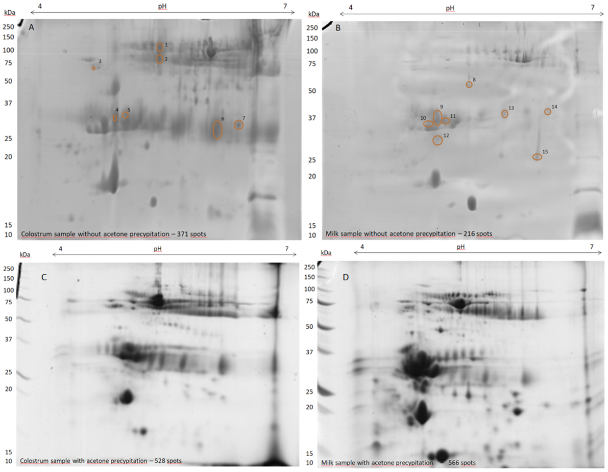
Fig. 1. Two-dimensional electrophoresis gels, stained with modified Bradford method, of mare colostrum (A) and milk (B) proteins without acetone precipitation and mare colostrum (C) and milk (D) with acetone precipitation; proteins (120 µg) were separated on 7 cm, 4–7 pH IPG strips; circles mark spots that have been cut out for identification using MALDI-TOF MS |
Rys. 1. Żele po dwukierunkowym rozdziale, wybarwione zmodyfikowaną metodą Bradforda, próbek siary (A) i mleka (B) klaczy, z pominięciem precypitacji acetonem i próby siary (C) i mleka (D) klaczy z precypitacją acetonem; białka (120 µg) zostały rozdzielone na 7 cm, 4–7 pH paskach IPG; okręgami zaznaczono spoty, które zostały wycięte w celu identyfikacji przy użyciu MALDI-TOF MS |
Isoelectric focusing on a linear, 7 cm pH 4–7 ReadyStrip IPG strips (Bio-Rad) applied to 120 µg of protein was carried out in two stages in PROTEAN® i12 IEF Cell (Bio-Rad). The first was passive rehydration (6h, 20°C), followed by active rehydration (12 h, 20°C, 50 V). The next stage was proper isoelectric focusing carried out according to the following procedure: 50 V for 100 Vh, 250 V for 150 Vh, 500 V for 500 Vh, 1000 V for 1000 Vh, 2 h for linear voltage increase from 1000 V to 5000 V, then 5000 V through 90000 Vh. After focusing, the strips were equilibrated for 15 min in a base buffer consisting of: 6 M urea, 0.5 M Tris/HCl o pH 6.8, 2% w/v SDS, 30% w/v glycerol with 1% DTT. After 15 min, the strips were transferred to the same base buffer with the addition of 2.5% iodoacetamide for 20 min. Electrophoresis was carried out at 40 V for 1 h and specific electrophoresis at 100 V for 90 min on small 12% polyacrylamide gels in Tris-glycine buffer (25 mM Tris-HCl, 192 mM glycine, 0.1% SDS) using Mini-PROTEAN® Tetra Cell (Bio-Rad). After completion of two-dimensional electrophoresis, gels were stained according to a modified Bradford method using Protein Assay Dye Reagent Concentrate (Bio-Rad) mixed v/v 1:20 with super pure water.
Stained gels were scanned using a GS-800TM Calibrated Densitometer (Bio-Rad). Two-dimensional images (2-D gel) were processed and subjected to quantitative and qualitative analysis using PDQuest Advanced Analysis Software 8.0.1 advanced (Bio-Rad).
After cutting out the imaged protein spots, an attempt was made to identify them using a MALDI-TOF mass spectrometer.
Before electrophoretic separation is performed, the samples must be dissolved in a lysis buffer (necessary to keep proteins dissolved and unbound during proteomic analyses during for a separation of proteins on polyacrylamide gels). The first step in preparing samples for proteomic analyses is their precipitation using e.g. acetone. In the prepare of certain tissues, e.g. the brain, it is necessary to use acetone precipitation due to the low loss of protein during sample preparation [Fic et al. 2010Fic, E., Kedracka-Krok, S., Jankowska, U., Pirog A., Dziedzicka-Wasylewska, M. (2010). Comparison of protein precipitation methods for various rat brain structures prior to proteomic analysis. Electrophoresis, 31(21), 3573–3579. https://doi.org/10.1002/elps.201000197]. The aim of precipitation is to obtain proteins from the sample and to remove the compounds interfering with 2-D electrophoresis. Due to the difficulty of dissolving in lysis buffer samples of mare colostrum and milk precipitated in acetone in the present study were checked whether this step can be skipped when preparing samples for 2-DE separation.
Images of 2-DE gels of mare colostrum (A) and milk (B) which not have been precipitated and for comparison images of 2-DE gels of colostrum (C) and milk (D) that were subjected to acetone precipitation are shown in Fig. 1.
Bioinformatic analysis using PDQuest Advanced Analysis Software 8.0.1 advanced (Bio-Rad) showed on the 2-DE gel 371 colostrum protein spots and 216 milk protein spots. The stained protein spots on 2-DE images were smudged and the obtained image resolution was very low. The result was the loss of many protein spots. Omission of the acetone precipitation of the samples also prevented from the identification of the separated proteins using a MALDI-TOF spectrometer. For comparison, as a result of bioinformatic analysis of 2-DE mare colostrum and milk gels after acetone precipitation shown that 528 spots were on colostrum 2-DE gel and 566 spots on milk 2-DE gel.
Proteomic analyses allow the determination of the entire protein composition of a given biological material (organs, tissues, body fluids). Various research tools are used to determine the proteome of body fluids. For example, 1-DE or 2-DE and LC-MS/MS, MALDI-TOF MS, nanoLC LTQ Orbitrap MS or gel and liquid chromatography/quadrupole [Bereman et al. 2009Bereman, M., Williams, T., Muddiman, C. (2009). Development of nanoLC LTQ Orbitrap Mass spectrometric Method for Profiling Glycans Derived from Plasma from Healthy, Benign Tumor Control, and Epithelial Ovarian Cancer Patients. Anal. Chem., 81(3), 1130–1136. https://doi.org/10.1021/ac802262w, Sandra et al. 2010Sandra, K., Pereira, A., Vanhoenacker, G., David, F., Sandra, P. (2010). Comperehensive blood plasma lipidomics by liquid chromatography/quadrupole time-of-flight mass spectrometry. J. Chromatogr., 1217, 4087–4099. https://doi.org/10.1016/j.chroma.2010.02.039, Kalra et al. 2013Kalra, H., Adda, Ch., Liem, M., Ang, Ch., Mechler, A., Simpson, R., Hulett M., Mathivanan, S. (2013). Comparative proteomics evaluation of plasma exosome isolation techniques and assessment of the stability of exosomes in normal human blood plasma. Proteomics, 13(3), 3354–3364. https://doi.org/10.1002/pmic.201300282, Dratwa-Chałupnik et al. 2016aDratwa-Chałupnik, A., Ożgo, M., Herosimczyk, A., Lepczyński, A., Michałek, K. (2016a). Lactose-rich milk replacer mofifies the promote of blood plasma in 2-week-old calves. Turk. J. Vet. Anim. Sci., 40, 21–27. https://doi.org/10.3906/vet-1504-57, 2016b, Lepczyński et al. 2017Lepczyński, A., Ożgo, M., Dratwa-Chałupnik, A., Robak, P., Pyć, A., Zaborski, D., Herosimczyk, A. (2017). An update on medium- and low-abundant blook plasma proteome of horse. Animal, 1, 1–12. https://doi.org/10.1017/S1751731117001409]. So far, the protein profile of milk has been determined primarily for ruminants using for this purpose 1-DE in combination with LC-MS/MS [Ha et al. 2015Ha, M., Sabherwal, M., Duncan, E., Stevens, S., Stockwell, P., McConnell, M., Bekhit, A., Carne, A. (2015). In-Depth Characterization of Sheep (Ovis aries) Milk Whey Proteome and Comparision with Cow (Bos taurus). PLoS ONE 10(10): e0139774. https://doi.org/10.1371/journal.pone.0139774], 2-DE in combination with MALDI-TOF MS [Di Girolamo et al. 2014Di Girolamo, F., Masoti, A., Salvatori, G., Scapaticci, M., Muraca, M., Putignani, L. (2014). A sensitive and efective proteomic approach to identify she-donkey's and goat's milk adulterations by MALDI-TOF MS ingerprinting. Int. J. Mol. Sci., 15, 13697–13719. https://doi.org/10.3390/ijms150813697], 2DE/ELISA [Hernández-Castellano et al. 2016Hernández-Castellano, L., Almeida, A., Renaut, J., Argüello, A., Castro, N. (2016). A proteomics study of colostrum and milk from the two major small ruminant dairy breeds from the Canary Islands: A bovine milk comparison perspective. J. Dairy. Res., 83, 366–374. https://doi.org/10.1017/S0022029916000273] and affinity chromatography and nLC-Chip-QTOF-MS [Le Parc et al. 2014Le Parc, A., Dallas, D., Duaut, S., Leonil, J., Martin, P., Barile, D. (2014). Characterization of goat milk lactoferrin N-glycans and comparison with the N-glycomes of human and bovine milk. Electrophoresis, 35(11), 1560–1570. https://doi.org/10.1002/elps.201300619].
Depending on the equipment used for proteomic analyses, the method of sample preparation varies. Our previous research developed a preliminary methodology for preparing mare colostrum and milk samples for proteomic analyses using 2-D electrophoresis in combination with MALDI-TOF mass spectrometry (unpublished materials, research project for young scientists no. 8230). According to the developed methodology, colostrum and milk samples were first skimmed, then caseins were removed from them, and in the next stage they were precipitated with acetone. The prepared samples were dissolved in lysis buffer. The process of dissolving acetone-precipitated samples in lysis buffer was lengthy and difficult, especially for mare colostrum samples. Therefore, the presented studies omitted the stages of acetone precipitation, which increased and accelerated the process of dissolution of samples in the lysis buffer compared to samples that have previously been precipitated with acetone. Unfortunately, skipping the precipitation stage with acetone resulted in blurry images of 2-DE gels. Most likely, the reason for this was the presence of interfering compounds in the 2-DE separation.
Apart from the precipitation of proteins, acetone’s task is to reduce the amount of interfering compounds mainly salts. In the case of blood plasma/serum, it is sufficient to clean them of non-protein components by precipitation with acetone [Herosimczyk et al. 2013Herosimczyk, A., Lepczyński, A., Dratwa-Chałupnik, A., Michałek, K., Skrzypczak, W. (2013). Blood plasma protein and lipid profile changes in calves during the first week of life. Pol. J. Vet. Sci., 16(3), 425–434. https://doi.org/10.2478/pjvs-2013-0060, Dratwa-Chałupnik et al. 2016aDratwa-Chałupnik, A., Ożgo, M., Herosimczyk, A., Lepczyński, A., Michałek, K. (2016a). Lactose-rich milk replacer mofifies the promote of blood plasma in 2-week-old calves. Turk. J. Vet. Anim. Sci., 40, 21–27. https://doi.org/10.3906/vet-1504-57, Lepczyński et al. 2017Lepczyński, A., Ożgo, M., Dratwa-Chałupnik, A., Robak, P., Pyć, A., Zaborski, D., Herosimczyk, A. (2017). An update on medium- and low-abundant blook plasma proteome of horse. Animal, 1, 1–12. https://doi.org/10.1017/S1751731117001409, Gondek et al. 2019Gondek, M., Herosimczyk, A., Knysz, P., Ożgo, M., Lepczyński, A., Szkucik, K. (2019). Comparative Proteomic Analysis of Serum from Pig Experimentally Infected with Trichinella spiralis, Trichinella britovi, and Trichinella pseudospiralis. Patchogens, 9(55), 1–28. https://doi.org/10.3390/pathogens9010055]. Preparation of cerebrospinal fluid samples for proteomic analyses requires concentration and precipitation with acetone to remove non-protein components [D'Aguanno et al. 2008D'Aguanno, S., Barassi, A., Lupisella, S., Melzi d'eril, G., Del Boccio, P., Pieragostino, D., Pallotti, F., Carelli, V., Valentino, M., Liguori, R., Anoni, P., Bernardini, S., Gambi, D., Urbani, A., Federici, G. (2008). Differential cerebro spinal fluid proteome investigation of Leber hereditary optic neuropathy (LHON) and multiple sclerosis. J. Neuroim., 193, 156–160. https://doi.org/10.1016/j.jneuroim.2007.10.004]. When preparing urine samples for proteomic analyses, purifying samples with acetone is not enough. For proteomic analysis of urine of calves using 2-DE and MS, the samples had to be desalted with a Clean Up Kit [Dratwa-Chałupnik et al. 2016bDratwa-Chałupnik, A., Ożgo, M., Lepczyński, A., Herosimczyk, A., Michałek, K. (2016b). Excessive amount of lactose in the diet of two-week-old calves induces urinary protein changes. Arch. Anim. Breed., 59, 417–422. https://doi.org/10.5194/aab-59-417-2016]. In contrast, saliva samples are directly dissolved in lysis buffer without precipitation of acetone [Baldini et al. 2011Baldini, C., Giusti, L., Ciregia, F., Da Valle, Y., Giacomelli, C., Donadio, E., Sernissi, F., Bazzichi, L., Giannaccini, G., Bombardieri, S., Lucacchini, A. (2011). Proteomic analysis of saliva: a unique tool to distinguish primary Sjorgen's syndrome from secondary Sjorgen's syndrome and other sicca syndromes. Arthritis Res. Ther., 13, 2–16. https://doi.org/10.1186/ar3523].
The composition of colostrum and milk varies in species, so a sample preparation procedure dedicated to a specific animal species should be developed. Golinelli et al. [2011]Golinelli, L., Conte-Junior, C., Paschoalin, V., Silva, J. (2011). Proteomic Analysis of Whey from Bovine Colostrum and Mature Milk. Braz. Arch. Biol. Technol., 54(4), 761–768. https://doi.org/10.1590/S1516-89132011000400016, developing a methodology for preparing colostrum and cow milk samples for proteomic analyses using 2-DE and MS, compared two research methods. The first method used the Clean Up Kit, the second was based on the precipitation of samples with 10% trichloroacetic acid (TCA). The authors show that samples subjected to precipitation by TCA gave two-dimensional map with better resolution and clearest background compared with Clean Up Kit treatment.
In the presented work, omitting the precipitation of acetone samples resulted in better solubility of mare milk and colostrum in the lysis buffer, but adversely affected the 2-D electrophoretic image. In addition, contamination of the sample with non-protein compounds prevented the identification of protein spots using MALDI-TOF MS. Perhaps to purify the sample from interfering compounds, a more effective method than precipitation with cold acetone would be precipitation with trichloroacetic acid. To this end, further research is needed to optimize the procedure for preparing colostrum and mare milk samples for proteomic analyses using 2-DE and MS.
According to the available literature, these proteomic studies of mare’s colostrum and milk based on 2-DE and MALDI-TOF MS are the first undertaken to this topic.
Preparation of mare’s colostrum and milk sample without samples precipitation of acetone did not bring the assumed results. Substances, including salts, witch interfering with electrophoretic separation, as a result of which the 2-DE image stained protein spots were smudged, and the obtained image resolution was very low. At the same time, salt contamination of the samples prevented from identification of the obtained protein spots using a MALDI-TOF MS.
Therefore, the procedure for preparing mare colostrum and milk samples by the stage of purifying samples from non-protein substances simultaneously maintaining solubility proteins in the lysis buffer should be worked out. The methodology for preparing mare colostrum/milk samples for proteomic analyses described in this work can be helpful in developing the optimal procedure for preparing these samples for proteomic analyses using 2-DE and MS.
Research was financed from funds for young researchers (project number 8230).
Received: 22 Jul 2020
Accepted: 18 Sep 2020
Published online: 2 Oct 2020
Accesses: 917
Cichy, A., Dratwa-Chałupnik, A., Medeńska, W., Ożgo, M., Pikuła, R., (2020). Preparation of mare's colostrum and milk sample for 2-DE separation without acetone precipitation. Acta Sci. Pol. Zootechnica, 19(3), 71–78. DOI: 10.21005/asp.2020.19.3.09.