Case Study
Dominika M. Karasiewicz ![]() 1 , Anna Kołodziejska-Sawerska
1 , Anna Kołodziejska-Sawerska ![]() , Magdalena Stanek
, Magdalena Stanek ![]()
Department of Animal Physiology and Physiotherapy, Faculty of Animal Breeding and Biology, Bydgoszcz University of Science and Technology, Prof. Sylwestra Kaliskiego 7, 85-796 Bydgoszcz, Poland
Abstract. The aim of this study was to determine the effectiveness of a rehabilitation program during recovery from surgery in a dog with patellar dislocation and rupture of the cranial cruciate ligament. Manual techniques, active and passive kinesitherapy and hydrotherapy were used in the study. Parametric methods were used to evaluate the effectiveness of physiotherapy: goniometric measurements, lameness severity analysis, pelvic limb circumference measurements, and a non-parametric method: a questionnaire. The results of our study showed that physiotherapy had a positive effect on restoring normal gait and increasing muscle mass in the operated limb. The study confirmed the great usefulness of rehabilitation in the recovery process after surgical stabilization of knee joint structures. Beginning physiotherapy as soon as possible after the surgery is crucial to prevent the negative effects of limb immobilization.
Keywords: animal physiotherapy, cranial ligament rupture, patellar luxation, hydrotherapy, dog
The cranial cruciate ligament is the most important anatomical structure stabilizing the knee joint [Aleksiewicz et al. 2011Aleksiewicz, R., Niedziela, D., Cichecki, M., Bojarski, M. (2011). Zastosowanie ligatury Codoloops kotwioneJ. wszczepami drutu chirurgicznego w zabiegach naprawy zerwanego więzadła krzyżowego doczaszkowego u psa [Codoloops ligature application anchored with surgical wire implants in the reconstruction of cranial cruciate ligaments in dogs]. Med. Wet., 67(8), 550–554 [in Polish]. Google Scholar, Millis et al. 2016Millis, D.L., Levine, D., Taylor, R.A. (2016). Rehabilitacja psów [Canine rehabilitation]. Elsevier Urban & Partner, Wrocław [in Polish]. Google Scholar]. Its damage occurs due to degeneration developing with the age of the dog and as a result of trauma, the destructive forces of which significantly exceed the strength of the ligament. Other causes of damage to this structure are metabolic, nutritional, genetic diseases and abnormal anatomical structure related to the tibial plane angle (TPA). The only valid solution for ligament rupture is surgical treatment [Millis et al. 2016Millis, D.L., Levine, D., Taylor, R.A. (2016). Rehabilitacja psów [Canine rehabilitation]. Elsevier Urban & Partner, Wrocław [in Polish]. Google Scholar]. Many surgical techniques have been developed for stabilizing the ligament, which have been assigned to the following groups: intra-articular, extra-articular, and methods related to altering biomechanics in the knee joint through tibial tuberosity grafting.
The second leading cause of lameness in dogs is dislocation of the patella. This disorder leads to the development of a severe pain response, lameness, and degenerative disease of the knee joint [Johnson et al. 2001Johnson, A.L., Probst, C.W., Decamp, Ch.E., Rosenstein, D.S., Hauptman, J.G., Weaver, B. T., Kern, T.L. (2001). Comparison of trochlear block recession and trochlear wedge recession for canine patellar luxation using a cadaver model. Vet. Surg., 30, 140–150. https://doi.org/10.1053/jvet.2001.21391]. There are two types of patella dislocation: medial and lateral. Patellar dislocation to the medial side occurs in small and miniature breed dogs as opposed to lateral, affecting large and giant breed dogs [Di Dona et al. 2018Di Dona, F., Della Valle, G., Fatone, G. (2018). Pattelar luxation in dogs. Veterinary Medicine: Research and Reports, 9, 23–32. https://doi.org/10.2147/VMRR.S142545]. The causes leading to destabilization of this patella are related to nutritional, environmental, traumatic or genetic factors. Untreated dislocation of the patella is the cause of tearing or rupture of the medial collateral ligament, the occurrence of cartilage degeneration and the appearance of inflammation in the knee joint [Nganvongpanit et al. 2017Nganvongpanit, K., Buddhachat, K., Boonsri, B., Sripratak, T., Punyapornwithaya, V. (2017). Retrospective study of medial patellar luxation surgery using combination of four techniques without bone reconstruction in non-flattened femoral sulcus: 133 Cases in 10 Years' Period (2006–2015). Kafkas Uni. Vet. Fak. Derg., 23(2), 201 209. https://doi.org/10.9775/kvfd.2016.16218]. Conservative treatment is associated with the administration of supplements and anti-inflammatory drugs and rehabilitation. Surgical treatment involves stabilization of the patella and includes several surgical approaches.
The yorkshire terrier is a breed that is particularly predisposed to patella dislocation and rupture of the cranial cruciate ligament [Taylor-Brown et al. 2015Taylor-Brown, F.E., Meeson, R.L., Brodbelt, D,C., Church, D.B., McGreevy, P.D., Thomson, P.C., O'Neill, D.G. (2015). Epidemiology of Cranial Cruciate Ligament Disease Diagnosis in Dogs Attending Primary-Care Veterinary Practices in England. Veterinary Surgery, 44(6), 777–783. https://doi.org/10.1111/vsu.12349, O’Neill et al. 2016O’Neill, D.G., Meeson, R.L., Sheridan, A., Church, D.B., Brodbelt, D.C. (2016). The epidemiology of patellar luxation in dogs attending primary-care veterinary practices in England. Canine Genetics and Epidemiology, 4. https://doi.org/10.1186/s40575-016-0034-0]. Both conditions can occur simultaneously and lead to instability of the knee joint, muscle atrophy, pain, inflammation and stiffness of the joint resulting in limited range of motion. A number of diagnostic methods have been developed to allow early diagnosis of knee joint pathology. This allows for immediate surgical treatment to minimize the occurrence of severe musculoskeletal dysfunction. Surgical treatment seems to be the only correct method leading to a permanent stabilization of these anatomical structures.
Rehabilitation in both conditions is essential to restore motor function to the pelvic limb. Physiotherapy after surgical treatment of the damaged cruciate ligament and patella dislocation focuses primarily on postoperative pain relief, reduction of inflammation, restoration of full range of motion of the knee joint, re-education of active muscle stabilization and shaping proprioception in the knee joint. A key goal of rehabilitation is also to achieve loading of the limb, thereby restoring muscle mass [Chanoit and Sawaya 2007Chanoit, G., Sawaya, S.G. (2007). Physiotherapy and functional rehabilitation after cranial cruciate ligament surgery in dogs. Pratique Vet. [https://www.researchgate.net/publication/287056852\_Physiotherapy\_and\_functional\_rehabilitation\_after\_cranial\_cruciate\_ligament\_surgery\_in\_dogs] Google Scholar, Wąsiatycz and Wąsiatycz 2012Wąsiatycz, G., Wąsiatycz, R. (2012). Uszkodzenie więzadła krzyżowego przedniego stawu kolanowego u psów. Cz. III Rehabilitacja pooperacyjna uszkodzonego więzadła krzyżowego doczaszkowego stawu kolanowego [Cranial cruciate ligament injuries in canines. Part 3. Postoperative rehabilitation of the injured cranial cruciate ligament]. Weterynaria w Praktyce, 7–8, 19–33 [in Polish]. Google Scholar].
The aim of this study was to evaluate the effect of a rehabilitation program on the degree of lameness and changes in muscle mass and range of motion in the knee joint in a Yorkshire terrier dog after surgical stabilization of the patella and medial collateral ligament. The effect of physiotherapy was assessed on the basis of measurements of pelvic limb muscle circumference, range of motion in the knee joints, a lameness assessment card and a questionnaire.
Hypothesis: The use of massage, kinesitherapy and hydrotherapy after surgical treatment of patella dislocation and rupture of the cranial cruciate ligament allows to achieve the full range of motion at the knee joint, to increase the muscle mass of the pelvic limbs and to restore the normal gait pattern.
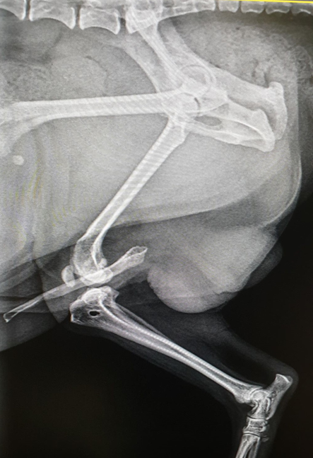
A Yorkshire terrier dog, aged 9 years, patient of the Centrum Veterinary Clinic in Bydgoszcz participated in the study. Patient was admitted to the clinic with lameness of the right pelvic limb and suspected rupture of the anterior cruciate ligament of the right knee joint. Clinical examination revealed decreased muscular contour of the right leg in comparison with the left pelvic limb. Observation of the dog's gait revealed unilateral grade 2/5 lameness of the right pelvic limb with toe rest and external rotation of the knee. During manipulation of the right knee, a partial intracranial drawer motion with a soft endpoint was noted. Additionally, a medial patellar dislocation of the fourth degree was noted on orthopedic examination. X-rays of the right knee joint taken in medial-lateral and horizontal projections showed a tear of the medial collateral ligament and a medial dislocation of the patella. Based on the clinical and orthopedic examination, a provisional diagnosis of patella dislocation and right anterior cruciate ligament rupture was established.
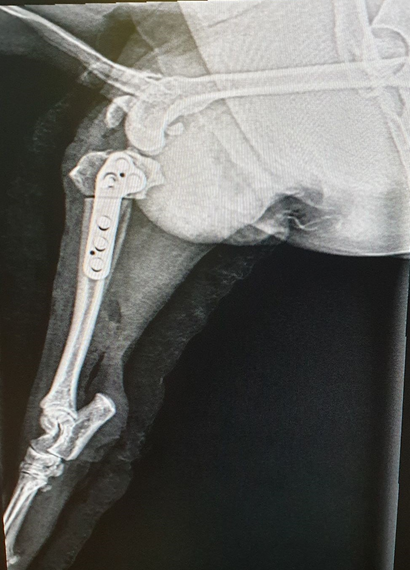
Due to postoperative complications the patient underwent 3 surgical procedures for stabilization of the ruptured cranial cruciate ligament of the right pelvic limb. Extracapsular stabilization (side-to-side suture), intracapsular stabilization (synthetic implant), and a method to prevent tibial slippage in the intracapsular direction (TTA-Rapid) were performed. Additionally, surgical repositioning of the patella was performed using the femoral block deepening technique. Four weeks after the last surgical procedure, the patient was referred to the rehabilitation due to lameness and significant muscle atrophy. During the rehabilitation program measures were taken to improve muscles in the operated pelvic limb, to relax tight muscles in both pelvic limbs and to improve the range of motion in the pelvic limb after stabilization of the cruciate ligament and patella.
\bigskip
|
Fig. 1. X-ray picture of the patient before the procedure (Own material) |
Manual therapy consisting of massage was used to begin each therapeutic session. Stroking was used to initiate and intertwine the various massage modalities and to conclude the manual therapy. The second massage technique was shallow and deep rubbing. It was followed by shallow and deep kneading, and circular compression and broomstick. The next step was shaking the muscle parts and limbs. The manual therapy lasted 20 minutes. After the massage, passive range of motion (PROM) exercises were performed in the joints of the pelvic limbs with stretching elements. 30 repetitions were performed in each joint of the pelvic limbs. The following types of passive movements were used: flexion, extension, inversion, adduction and rotation.
The next form of rehabilitation was active kinesitherapy. The training plan included balancing, snoppy exercise, sit-stand, manual limb relief in standing position, walking on an uneven surface, active stretching, and dancing. The exercises included were tailored to the animal's fitness level and modified over time. Sensory discs, Donut and Peanut balls, Cavaletti obstacles, uneven surface mat, inclined surface, steps and slalom were used during the course.
Hydrotherapy was an important element of the rehabilitation procedure, which contributed to the development of muscle mass and improved the range of motion of the right knee joint. It was carried out with the use of a HYCON water treadmill type MACBD. The workouts lasted 20 minutes. Exercises in the water treadmill were introduced from the first rehabilitation classes always after the massage. The water temperature during the session was 24° C and the water level reached the patient's shoulder joint. No countercurrent was used during the rehabilitation procedure. The speed of the treadmill belt movement was adjusted to the animal's capabilities and constantly controlled.
Progress monitoring during rehabilitation was performed using the following research tools: measurement of pelvic limb circumferences, measurement of the range of motion in the knee joint of both pelvic limbs, lameness rating scale and questionnaire. The first parameter measured was the circumference of the muscle mass of the pelvic limbs. It was performed in two places on both pelvic limbs using a measure, indicating the result with an accuracy of 1 mm. During the measurements, the dog was in a recumbent position. The first measurement was taken above the knee joint at mid-thigh, while the second measurement was taken below the knee joint at mid-shank. Measurements were taken three times at each location to obtain a reliable result.
The next step was to measure the range of motion of the knee joint of the right and left pelvic limb. A SAEHAN goniometer was used to measure this parameter. The passive range of motion of the joint during maximal extension and flexion of the limb in the knee joint was measured. Each test was repeated three times to obtain a reliable result. The measurements obtained were compared with normative values based on literature data, which assume 40–45° for flexion at the knee joint and 160–170° for extension [Millis et al. 2016Millis, D.L., Levine, D., Taylor, R.A. (2016). Rehabilitacja psów [Canine rehabilitation]. Elsevier Urban & Partner, Wrocław [in Polish]. Google Scholar, Levine et al. 2017Levine, D., Bockstahler, B., Milis, D. (2017). Fizjoterapia psów i kotów. Rehabilitacja i zwalczanie bólu [Physiotherapy for dogs and cats. Rehabilitation and pain relief]. Galaktyka, Łódź [in Polish]. Google Scholar]. Measurements were taken before and after the rehabilitation session.
The severity of lameness was assessed by observing the way the animal moved at trot and trot, at the first and last rehabilitation session. The results of the gait analysis were then compared with Table 1, which includes lameness scores. At the end of rehabilitation, the owner completed a questionnaire collecting information on the effectiveness of the rehabilitation program applied, relating to the animal's daily functioning and activity. Sixteen rehabilitation encounters were implemented. They took place three times a week.
|
Fig. 2. X-ray picture of the patient after using the TTA-Rapid technique (Own material) |
Table 1. Degrees of severity of lameness [Milis et al. 2014] |
|
Lameness score |
Description |
0 |
Unchanged state |
1 |
Slight, intermittent lameness |
2 |
Marked lameness with pelvic limb loading |
3 |
Severe lameness with pelvic limb strain |
4 |
Intermittent lameness without weight on the pelvic limb |
5 |
Continued lameness without putting weight on the pelvic limb |
To evaluate the results obtained from the pelvic limb circumference and goniometric measurements of the knee joint of both limbs, the collected results were compiled in Excel, where the arithmetic mean of the three measurements taken at each rehabilitation session was calculated. Then, after completing the physiotherapy program, Student's t-test was performed for samples with the dependent variable using Statistica 13.1. PL program. Significance of the differences in the mean values of the measurements before and after therapy session were determined at for P ≤ 0.05. The statistical analysis of muscle measurements and range of motion in the joints concerned the same part of the limb, for example the left and right sides before rehabilitation and the left and right sides after rehabilitation.
Table 2. shows the results of the measurements of the right and left pelvic limb circumferences. The circumferences within each pelvic limb differed statistically significantly before and after the physiotherapeutic management. This is due to the positive effect of rehabilitation management on the increase of muscle mass. Comparing the final values of the measurements of the right pelvic limb affected by the injury in relation to the values of the left pelvic limb, uneven muscle development is found. The left pelvic limb was characterized by significantly greater gains in muscle mass. In the operated limb, the perimeters took a lower value and muscle growth was slower compared to the non-injured limb. During the physiotherapy program, equal values were not achieved in both pelvic limbs.
Table 3. shows the results of goniometric measurements of the knee joint in the healthy and surgically treated pelvic limb. Extension and flexion in the left knee joint before rehabilitation were within normal values. On the other hand, in the right pelvic limb, a significant restriction of movement was observed, which was related to the stiffness of the joint during both straightening and bending, caused by long-term disuse of the limb. The rehabilitation management applied contributed to statistically significant changes in the mobility of the right and left knee joint. The techniques used resulted in an increase in the angle during extension and flexion in the right limb to normative values.
The measurement results in Table 4. showing the two phases of the patient's movement (walk and trot) were for the right pelvic limb affected by the injury and subjected to surgical interventions. It shows that the rehabilitation program applied allowed the complete abolition of lameness in tarsus. However, a significant reduction to light and intermittent lameness was achieved at the trot. Before the physiotherapy treatment, the lameness in the dog's basic gait was intermittent with the limb not being loaded. At the trot, there was continuous lameness associated with not loading the limb.
The questionnaire completed by the caregiver indicated that the rehabilitation techniques used increased the animal's activity. The owner observed a reduction in lameness, which was associated with an improvement in the dog's comfort of living and willingness to undertake daily activities such as walks, playing with household members, going up and down stairs, and jumping on the couch.
Table 2. Measurement of the circumference of the healthy and operated pelvic limb, cm |
||||
|
Left side |
Right side |
||
before |
after |
before |
after |
|
Thigh |
12.0 |
18.0* |
10.3 |
12.0* |
Shank |
7.0 |
10.5* |
4.4 |
7.7* |
*means in the rows within one limb differed significantly (P < 0.05) before and after therapy. |
||||
Table 3. Measurement of the range of motion in the knee joint of the healthy and operated limb |
||||
|
Left side |
Right side |
||
before |
after |
before |
after |
|
Straighten |
163 |
168* |
138 |
169* |
Bend |
42 |
35* |
51 |
41* |
*means in the rows within one limb differed significantly (P < 0.05) before and after therapy. |
||||
Table 4. Evaluation of lameness before and after therapy |
||
|
Lameness score |
|
before |
after |
|
Walk |
4 |
0 |
Trot |
5 |
1 |
Degenerative changes leading to ligament rupture are slower in small dog breeds than in large or giant breeds. Therefore, this condition occurs in older dogs. The average age for small breed dogs in the work conducted by Dyall and Schmökel [2017]Dyall, B., Schmökel, H. (2017). Tibial tuberosity advancement in small-breed dogs using TTA Rapid implants: complications and outcome. J. Small Anim. Pract., 58, 314–322. https://doi.org/10.1111/jsap.12654 was about 8 years. Wąsiatycz [2010]Wąsiatycz, G. (2010). Uszkodzenie więzadła krzyżowego przedniego stawu kolanowego u psów Cz. I Najpopularniejsze techniki operacyjne [Cranial cruciate ligament rupture in dogs. Part 1. The most popular surgical techniques]. Weterynaria w Praktyce, 7(10), 10–19 [in Polish]. Google Scholar describes as consequences of damage to the intracranial cruciate ligament the occurrence of instability in the knee joint, which contributes to the development of osteoarthritis. In addition, pain is noticed, as well as atrophy of the thigh muscles and weakening of the strength of these muscles. The second main cause causing pelvic limb lameness is patella dislocation. Sterna et al. [2017]Sterna, J., Migdalska, A., Tomkowicz, A., Frymus, J., Degórska, B., Trębacz, P., Galanty, M. (2017). Praktyczne aspekty zwichnięcia rzepki u psów [Practical aspects of patella luxation treatment in dogs]. Życie Weterynaryjne, 92(8), 571–575 [in Polish]. Google Scholar in his work emphasizes that in small and miniature breeds, dislocation of the patella to the medial side occurs. In contrast, one of the main types of patients with patellar dislocation are older dogs. Often within this patient group, destabilization of the patella is accompanied by rupture of the intracranial cruciate ligament [Sterna et al. 2017Sterna, J., Migdalska, A., Tomkowicz, A., Frymus, J., Degórska, B., Trębacz, P., Galanty, M. (2017). Praktyczne aspekty zwichnięcia rzepki u psów [Practical aspects of patella luxation treatment in dogs]. Życie Weterynaryjne, 92(8), 571–575 [in Polish]. Google Scholar]. Yorkshire terrier dogs are predisposed to patella dislocation due to, among other things, shallowing of the femoral groove. This condition is accompanied by lameness and reduced range of motion in the knee joint [Lik 2016Lik, M. (2016). Zastosowanie terapii manualnych w rehabilitacji stawu kolanowego psa [The use of manual therapies in the rehabilitation of the canine knee joint]. https://www.researchgate.net/publication/324080702 [in Polish]. Google Scholar]. This is confirmed by the case of a patient who underwent rehabilitation treatment.
Wąsiatycz and Wąsiatycz [2012]Wąsiatycz, G., Wąsiatycz, R. (2012). Uszkodzenie więzadła krzyżowego przedniego stawu kolanowego u psów. Cz. III Rehabilitacja pooperacyjna uszkodzonego więzadła krzyżowego doczaszkowego stawu kolanowego [Cranial cruciate ligament injuries in canines. Part 3. Postoperative rehabilitation of the injured cranial cruciate ligament]. Weterynaria w Praktyce, 7–8, 19–33 [in Polish]. Google Scholar emphasize the role of physiotherapy in the recovery of the operated limb and intensive rehabilitation procedures applied in a short interval after surgery result in a faster and much more effective recovery of motor function of the injured limb. Moreover, recommendations applied a few years ago, consisting in limiting the animal's activity and immobilizing the limb for a few weeks, contributed to permanent pathological changes within the operated joint. They prevented the animal from returning to full fitness. Berte et al. [2012]Berte, L., Mazzanti, A., Salbego, F.Z., Beckamn, D.V., Santos R.P., Polidoro D., Baumhardt R. (2012). Immediate physical therapy in dogs with rupture of the cranial cruciate ligament submitted to extracapsular surgical stabilization. Arq. Bras. Med. Vet. Zoo., 64(1), 1–8. https://doi.org/10.1590/S0102-09352012000100001 indicated that rehabilitation should start from the first day after surgery. This makes it possible to prevent contractures and thus muscle atrophy. Also, Marsolais et al. [2002]Marsolais, G.S., Dvorak, G., Michael, G. (2002). Effects of postoperative rehabilitation on limb function after cranial cruciate ligament repait in dogs. J. Am. Vet. Med. Assoc., 220(9), 1325–1330. https://doi.org/10.2460/javma.2002.220.1325 studying the effect of early rehabilitation on the restoration of functional limb function after surgery for ruptured intracranial cruciate ligament concluded that physiotherapy should be an indispensable part of postoperative management. Their study showed that the difference between the function of the healthy limb and the operated limb in animals that had limited exercise after surgery was significant. This relationship can also be seen in the case of an individual who came to the Zoophysiotherapy Laboratory nearly 4 weeks after his last surgery. At the end of rehabilitation in our study, muscle mass gains were observed in both pelvic limbs, but the circumferences of the operated limb were smaller compared to the healthy limb. Berte et al. [2012]Berte, L., Mazzanti, A., Salbego, F.Z., Beckamn, D.V., Santos R.P., Polidoro D., Baumhardt R. (2012). Immediate physical therapy in dogs with rupture of the cranial cruciate ligament submitted to extracapsular surgical stabilization. Arq. Bras. Med. Vet. Zoo., 64(1), 1–8. https://doi.org/10.1590/S0102-09352012000100001 and Millis and Ciuperca [2015]Millis, D.L., Ciuperca, A. (2015). Evidence for canine rehabilitation and physical therapy. Vet. Clin. N Am-Small Anim., 45, 1–27. https://doi.org/10.1016/j.cvsm.2014.09.001 also emphasize the important impact of early application of rehabilitation, whereby it is possible to limit the worsening of muscle atrophy in the thigh and lower leg while rapidly rebuilding muscle mass and strength. In addition, such action allows for rapid achievement of full range of motion in the joints and prevents muscle contractures.
Wąsiatycz and Wąsiatycz [2012]Wąsiatycz, G., Wąsiatycz, R. (2012). Uszkodzenie więzadła krzyżowego przedniego stawu kolanowego u psów. Cz. III Rehabilitacja pooperacyjna uszkodzonego więzadła krzyżowego doczaszkowego stawu kolanowego [Cranial cruciate ligament injuries in canines. Part 3. Postoperative rehabilitation of the injured cranial cruciate ligament]. Weterynaria w Praktyce, 7–8, 19–33 [in Polish]. Google Scholar as methods used in rehabilitation after surgical treatment of the cruciate cranial ligament indicate: kinesitherapy (PROM, slow walking, walking on an inclined surface and steps, water treadmill, sit-stand exercise, Cavaletti obstacles, dance, walking on sand), massage, heat therapy, cryotherapy, electrical stimulation, ultrasound. Chanoit and Sawaya [2007]Chanoit, G., Sawaya, S.G. (2007). Physiotherapy and functional rehabilitation after cranial cruciate ligament surgery in dogs. Pratique Vet. [https://www.researchgate.net/publication/287056852\_Physiotherapy\_and\_functional\_rehabilitation\_after\_cranial\_cruciate\_ligament\_surgery\_in\_dogs] Google Scholar also recommends the use of massage, passive joint motion exercises, electrostimulation, cryotherapy, heat therapy, ultrasound, hydrotherapy, stretching, slow walking on a flat and uneven surface, manual limb decompression, exercises referred to as dancing and sit-ups, inclined surfaces and walking steps, and exercises using sensory cushions. Lemos et al. [2009]Lemos, C.M., Fischer, C.D.B., Baja, K.G., Pinto, V.M., Maia, J. (2009). World Small Animal Veterinary Association World Congress Proceedings – ``Physiotherapy Use in Rehabilitation of Dogs with Patella Luxation'', Sao Paulo, Brazylia. 21-24 lipca 2009. [https://www.vin.com/apputil/content/defaultadv1.aspx?pId=11290&id=4252857] Google Scholar emphasize that there are differences in rehabilitation programs for patients depending on the degree of patellar dislocation. They mention the following methods as helpful in the elimination of limb dysfunction caused by this condition: passive joint range of motion exercises, stretching, cryotherapy, electrostimulation, laser therapy, ultrasound and active kinesitherapy, acupuncture, hydrotherapy. The use of specific techniques depends primarily on the time that has passed since the operation. In the initial stage, the aim is to reduce pain, eliminate swelling, maintain the range of motion in the joint, eliminate tension in the muscles of the pelvic limbs, chest, back and neck. The elimination of swelling within the operated joint, reduction of inflammation, and acceleration of tissue healing are achieved through the use of: massage, lymphatic drainage, PROM passive exercises, cryotherapy, TENS, laser therapy, sonotherapy, acupuncture, thermotherapy, hydrotherapy, bicycle, stretching, and retraction reflex stimulation [Lemos et al. 2009Lemos, C.M., Fischer, C.D.B., Baja, K.G., Pinto, V.M., Maia, J. (2009). World Small Animal Veterinary Association World Congress Proceedings – ``Physiotherapy Use in Rehabilitation of Dogs with Patella Luxation'', Sao Paulo, Brazylia. 21-24 lipca 2009. [https://www.vin.com/apputil/content/defaultadv1.aspx?pId=11290&id=4252857] Google Scholar, Berte et al. 2012Berte, L., Mazzanti, A., Salbego, F.Z., Beckamn, D.V., Santos R.P., Polidoro D., Baumhardt R. (2012). Immediate physical therapy in dogs with rupture of the cranial cruciate ligament submitted to extracapsular surgical stabilization. Arq. Bras. Med. Vet. Zoo., 64(1), 1–8. https://doi.org/10.1590/S0102-09352012000100001, Lik 2016Lik, M. (2016). Zastosowanie terapii manualnych w rehabilitacji stawu kolanowego psa [The use of manual therapies in the rehabilitation of the canine knee joint]. https://www.researchgate.net/publication/324080702 [in Polish]. Google Scholar, Levine et al. 2017Levine, D., Bockstahler, B., Milis, D. (2017). Fizjoterapia psów i kotów. Rehabilitacja i zwalczanie bólu [Physiotherapy for dogs and cats. Rehabilitation and pain relief]. Galaktyka, Łódź [in Polish]. Google Scholar]. In the final stage of surgical care, elements of active kinesitherapy, passive kinesitherapy, and hydrotherapy are used [Levine et al. 2017Levine, D., Bockstahler, B., Milis, D. (2017). Fizjoterapia psów i kotów. Rehabilitacja i zwalczanie bólu [Physiotherapy for dogs and cats. Rehabilitation and pain relief]. Galaktyka, Łódź [in Polish]. Google Scholar]. The work performed included active and passive exercises, manual techniques, and hydrotherapy using a water treadmill at each session.
In our study, the patient showed significant atrophy of muscle mass in the operated right pelvic limb. The rehabilitation management performed allowed the patient to expand his muscle mass. However, the muscle growth was not uniform; a significantly slower development was observed in the injured limb. This was indicative of advanced atrophy and significant limitation of the range of motion and muscle tensions, which had to be eliminated. The development of muscle mass was mainly influenced by the applied active kinesiotherapy with the use of physiotherapy equipment and a water treadmill. Hydrotherapy resulted in a statistically significant increase in thigh and shin circumference values in the pelvic limb undergoing surgical intervention. Levine et al. [2017]Levine, D., Bockstahler, B., Milis, D. (2017). Fizjoterapia psów i kotów. Rehabilitacja i zwalczanie bólu [Physiotherapy for dogs and cats. Rehabilitation and pain relief]. Galaktyka, Łódź [in Polish]. Google Scholar highlight that water characteristics such as viscosity and resistance significantly contribute to muscle mass development.
Goniometric measurements prior to the rehabilitation procedure, confirmed normal values were found during extension and flexion of the left knee joint. However, in the right pelvic limb, a significant restriction of movement was noted due to stiffness of the joint during the extension and flexion phases. This was caused by long-term disuse of the limb due to postoperative complications. The application of physiotherapy treatments was effective and contributed to the improvement of flexion and extension angles in the right knee joint. According to Monk et al. [2006]Monk, M.L., Preaton, C.A., McGowan, C.M. (2006). Effects of early intensive postoperative physiotherapy on limb function after tibial plateau leveling osteotomy in dogs with deficiency of the cranial cruciate ligament. Am. J. Vet. Res., 67(3), 529–536. https://doi.org/10.2460/ajvr.67.3.529, Wąsiatycz and Wąsiatycz [2012]Wąsiatycz, G., Wąsiatycz, R. (2012). Uszkodzenie więzadła krzyżowego przedniego stawu kolanowego u psów. Cz. III Rehabilitacja pooperacyjna uszkodzonego więzadła krzyżowego doczaszkowego stawu kolanowego [Cranial cruciate ligament injuries in canines. Part 3. Postoperative rehabilitation of the injured cranial cruciate ligament]. Weterynaria w Praktyce, 7–8, 19–33 [in Polish]. Google Scholar, and Zink and van Dyke [2013]Zink, Ch.M., van Dyke, J.B. (2013). Canine sports medicine and rehabilitation. John Wiley & Sons Inc, Oxford. https://doi.org/10.1002/9781118783443, the following physiotherapeutic methods have a beneficial effect on improving joint range of motion: passive joint motion exercises combined with stretching, overcoming Cavaletti obstacles, hydrotherapy, walking on an inclined surface and steps, and active stretching using physiotherapy balls. In addition, Mikail [2006]Mikail, S. (2006). Hidroterapia. In: Fisioterapia Veterinária. Red. Mikail S., Pedro C. R., Manole, São Paulo, ss. 72-77 [in Portuguese]. Google Scholar confirms the positive effect of hydrotherapy on the range of motion in the knee joints, as well as the way the animal moves. Another study whose results correlate with our own regarding the positive effect of aquatic treadmill on goniometric measurements was the work of Preston and Wills [2018]Preston, T., Wills, A.P. (2018). A single hydrotherapy session increases range of motion and stride length in Labrador retrievers diagnosed with elbow dysplasia. Vet. J., 234, 105–110. https://doi.org/10.1016/j.tvjl.2018.02.013.
The study proved that rehabilitation using manual techniques, passive and active kinesiotherapy helped to reduce the degree of lameness. The physiotherapy applied allowed to completely eliminate lameness in the tarsus. A significant reduction to light and intermittent lameness was achieved in the trot. Scientific reports by Lik [2016]Lik, M. (2016). Zastosowanie terapii manualnych w rehabilitacji stawu kolanowego psa [The use of manual therapies in the rehabilitation of the canine knee joint]. https://www.researchgate.net/publication/324080702 [in Polish]. Google Scholar, Millis et al. [2016]Millis, D.L., Levine, D., Taylor, R.A. (2016). Rehabilitacja psów [Canine rehabilitation]. Elsevier Urban & Partner, Wrocław [in Polish]. Google Scholar and Levine et al. [2017]Levine, D., Bockstahler, B., Milis, D. (2017). Fizjoterapia psów i kotów. Rehabilitacja i zwalczanie bólu [Physiotherapy for dogs and cats. Rehabilitation and pain relief]. Galaktyka, Łódź [in Polish]. Google Scholar address rehabilitation programs that contribute to the elimination of lameness. These methods include slow walking, forcing weight on the affected limb, balancing on therapy balls, water treadmill, dance, Cavaletti obstacles, slalom, and walking on an uneven surface. The results of a study conducted by Innes et al. [2000]Innes, J.F., Bacon, D., Lynch, C., Pollard, A. (2000). Long-term outcome of surgery for dogs with cranial cruciate ligament deficiency. Vet. Rec., 147(12), 325–328. https://doi.org/10.1136/vr.147.12.325 confirm that properly conducted therapy in animals with damage to the medial collateral ligament has a positive effect on limp reduction. Monk et al. [2006]Monk, M.L., Preaton, C.A., McGowan, C.M. (2006). Effects of early intensive postoperative physiotherapy on limb function after tibial plateau leveling osteotomy in dogs with deficiency of the cranial cruciate ligament. Am. J. Vet. Res., 67(3), 529–536. https://doi.org/10.2460/ajvr.67.3.529 in their study found that active and passive kinesitherapy contributes to a reduction in lameness in dogs after surgical stabilization of the ligament.
Butterworth and Kydd [2017]Butterworth, S.J., Kydd, D.M. (2017). TTA-Rapid in the treatment of the canine cruciate deficient stifle: short- and medium-term outcome. J. Small Anim. Pract., 58, 35–41. https://doi.org/10.1111/jsap.12610 used a questionnaire to owners to verify the effectiveness of the physiotherapy performed and its impact on the comfort of the animal. Through it, they obtained independent results confirming the effectiveness of the subject of their study. The non-parametric evaluation conducted in the framework of our own work after the application of the rehabilitation program had the same assumption. The questionnaire completed by the caregiver indicated that the rehabilitation techniques used increased the animal's activity. The owner observed a reduction in lameness, which was associated with an improvement in the animal's comfort of living and willingness to undertake daily activities such as walks, playing with household members, climbing and descending stairs, and jumping on the couch. This proves that the rehabilitation procedure allowed the animal to recover without any discomfort related to pain and lameness.
The combination of manual techniques and kinesitherapy improved the range of motion in the injured knee joint. The rehabilitation methods applied allowed for the development of muscle mass and the rehabilitation program enabled the injured limb to be loaded again. The applied rehabilitation management significantly contributed to the reduction of lameness caused by the patella and the medial collateral ligament injury. Physiotherapy increased the patient's activity related to minimizing lameness, which contributed to a significant improvement in quality of life. It is justified to continue the research on the role of rehabilitation in the elimination of limb dysfunction after complicated surgical treatment of patella dislocation and cruciate ligament rupture.
The research was financed from departmental sources.
Received: 20 Jan 2022
Accepted: 15 Mar 2022
Published online: 27 Aug 2022
Accesses: 753
Karasiewicz, D.M., Kołodziejska-Sawerska, A., Stanek, M., (2022). Rehabilitation procedure after surgical stabilization of the patella and cranial cruciate ligament in a dog – case study. Acta Sci. Pol. Zootechnica, 21(1), 35–42. DOI: 10.21005/asp.2022.21.1.05.