Case Study
Sebastian Słodki ![]() 1,2, Joanna Bogucka
1,2, Joanna Bogucka ![]() 1
1
1Department of Animal Physiology and Physiotherapy, Bydgoszcz University of Science and Technology, Mazowiecka 28, 85-084 Bydgoszcz, Poland
2Sebastian Slodki Veterinary Office SweetVet, Łęczycka 37, 85-737 Bydgoszcz, Poland
Abstract. Rupture of the cranial cruciate ligament (CCL) in the stifle is one of the most common causes of hind-limb lameness in middle-aged and old dogs. The treatment of choice involves surgical extra- or intra-capsular stabilization of the knee joint, or the increasingly preferred tibial osteotomy techniques. Postoperative rehabilitation of the affected limb is an equally important part of the treatment. The paper describes three cases of rupture of the cruciate ligament in dogs and the methods of physiotherapeutic treatment applied to them. The techniques of passive and active exercises were used along with massage and physical therapy, such as magnetotherapy, ultrasound, electrotherapy, and laser therapy. The development of the correct physiotherapeutic treatment plan, the selection of techniques, and the duration of therapy is crucial for its effectiveness. It has been shown that rehabilitation has a positive effect on the tissue healing process. It also shortens the time needed to restore full mobility of the affected limb, both during the non-complicated healing process and in the event of problematic cases.
Keywords: Cranial cruciate ligament rupture, tibial osteotomy techniques, intra-capsular stabilization, dogs physiotherapy
The stifle is a compound joint that is formed by three bones: the distal part of the femur (os femur), the proximal part of the tibia (os tibia), and the largest sesamoid bone called the patella or knee cap [Millis and Levine 2013Millis, D., Levine, D. (2013). Canine Rehabilitation and Physical Therapy, Second Ed. Elsevier Saunders, Philadelphia, 1–760. Google Scholar]. Two joints can be distinguished in the knee: the femoral-tibial joint (articulatio femorotibialis) built by the femur, tibia, and menisci, and the femoral-patellar joint (articulatio femoropatellaris) [Done et al. 2009Done, S.H., Goody, P.C., Evans, S.A., Stickland, N.C. (2009). Color Atlas of Veterinary Anatomy, Volume 3, The Dog and Cat, Second Ed. Elsevier, 1–540. Google Scholar]. The stifle contains various types of tissues that must cooperate to maintain joint health and function [Cook 2010Cook, J.L. (2010). Cranial cruciate ligament disease in dogs: biology versus biomechanics. Vet. Surg., 39(3), 270–277. https://doi.org/10.1111/j.1532-950X.2010.00653.x]. There are four ligaments in the knee joint: the cranial cruciate ligament, the caudal cruciate ligament, and two collateral ligaments: medial and lateral [Canapp 2007Canapp, S.O. (2007). The Canine Stifle. Clin. Techn. Small Anim. Pract., 22(4), 195–205. https://doi.org/10.1053/j.ctsap.2007.09.008].
Stabilization of the knee during movement is possible thanks to the coordinated and synchronized work of the cranial cruciate ligament (CCL), the caudal cruciate ligament, the medial collateral ligament, the lateral collateral ligament, knee extension apparatus (consisting of the patellar ligament and quadriceps femoris), the gastrocnemius muscle, the biceps femoris [Adrian et al. 2013Adrian, C.P., Haussler, K.K., Kawcak, C., Reiser, R.F., Riegger-Krugh, C., Palmer, R.H., McIlwraith, C.W., Taylor, R.A. (2013). The role of muscle activation in cruciate disease. Vet. Surg., 42(7),765-773. https://doi.org/10.1111/j.1532-950X.2013.12045.x], the joint capsule, the synovial membrane, articular cartilage, and the lateral and medial meniscus [Cook 2010Cook, J.L. (2010). Cranial cruciate ligament disease in dogs: biology versus biomechanics. Vet. Surg., 39(3), 270–277. https://doi.org/10.1111/j.1532-950X.2010.00653.x].
A rupture of the cranial cruciate ligament is the most common cause of hind limb lameness in animals [Kirby 1993Kirby, B.M. (1993). Decision-making in cranial cruciate ligament ruptures. Vet. Clin. North Am. Small Anim. Pract., 23(4), 797–819. https://doi.org/10.1016/S0195-5616(93)50083-9, Bergh et al. 2014Bergh, M.S., Sullivan, C., Ferrell, C.L., Troy, J., Budsberg, S.C. (2014). Systematic Review of Surgical Treatments for Cranial Cruciate Ligament Disease in Dogs. J. Am. Anim. Hosp. Assoc., 50(5), 315–21. https://doi.org/10.5326/JAAHA-MS-6356]. The CCL has three main functions: to limit cranial displacement and internal rotation of the tibia, as well as to prevent knee hyperextension [Kim et al. 2010Kim, S.E., Pozzi, A., Banks, S.A., Conrad, B.P., Lewis, D.D. (2010). Effect of cranial cruciate ligament deficiency, tibial plateau leveling osteotomy, and tibial tuberosity advancement on contact mechanics and alignment of the stifle in flexion. Vet. Surg., 39(3), 363–370. https://doi.org/10.1111/j.1532-950X.2010.00655.x].
Because the pathogenesis of CCL rupture is quite complex, in order to systematise the subject the animals were divided into 4 groups. The first group were small dogs, mostly middle-aged, in whom CCL injury was secondary to dislocation of the patella to the medial side of the knee [Moore and Read 1996Moore, K.W., Read, R.A. (1996). Rupture of the cranial cruciate ligament in dogs. Part I. Compend. Contin. Educ. Pract. Vet., 18, 223–234. Google Scholar]. Moreover, many of these dogs were burdened with the defect of a gene, which was responsible for the development of femoral varus deformity, and this additionally burdened the CCL. The second group was medium and large dogs that had suffered a sudden injury. This damage led to significant hyperextension of the knee joint as a consequence of the strong compression of the CCL in the cranial direction relative to the intercondylar fossa, resulting in crushing and deformation of the ligament and ultimately its rupture [Slocum and Slocum 1993Slocum, B., Slocum, T.D. (1993). Tibial plateau leveling osteotomy for repair of cranial cruciate ligament rupture in the canine. Vet. Clin. North Am. Small Anim. Pract., 23, 777–95. https://doi.org/10.1016/S0195-5616(93)50082-7]. The third group were young, large dogs with congenital anatomical defects in the structure of the hind limbs, and above all with a tendency of hyperextension of the knee leading to continuous damage to the CCL, until its complete rupture [Slocum and Slocum 1993Slocum, B., Slocum, T.D. (1993). Tibial plateau leveling osteotomy for repair of cranial cruciate ligament rupture in the canine. Vet. Clin. North Am. Small Anim. Pract., 23, 777–95. https://doi.org/10.1016/S0195-5616(93)50082-7, Moore and Read 1996Moore, K.W., Read, R.A. (1996). Rupture of the cranial cruciate ligament in dogs. Part I. Compend. Contin. Educ. Pract. Vet., 18, 223–234. Google Scholar]. The fourth and the most numerous group were middle-aged and old dogs with chronic hind-limb lameness, as a consequence of osteoarthritis, most often with an immunological background [Vasseur 2003Vasseur, P.B. (2003). Stifle joint. In: Slatter D. (ed). Textbook of Small Animal Surgery, 3rd Ed. WB Saunders, Philadelphia, 2090–133. Google Scholar]. The chronic inflammatory and degenerative process, as well as pathological movement in the knee, led to gradual damage to the CCL, initially usually strain, caused by the structure of the CCL, and then to total rupture.
The diagnosis is based mainly on a physical examination, which usually detects swelling of the medial side of the knee joint, a positive cranial drawer sign, and a positive result of the tibial compression test [Kirby 1993Kirby, B.M. (1993). Decision-making in cranial cruciate ligament ruptures. Vet. Clin. North Am. Small Anim. Pract., 23(4), 797–819. https://doi.org/10.1016/S0195-5616(93)50083-9]. Other symptoms indicating rupture of the ligament are pain and muscular atrophies which appear later [Adrian et al. 2013Adrian, C.P., Haussler, K.K., Kawcak, C., Reiser, R.F., Riegger-Krugh, C., Palmer, R.H., McIlwraith, C.W., Taylor, R.A. (2013). The role of muscle activation in cruciate disease. Vet. Surg., 42(7),765-773. https://doi.org/10.1111/j.1532-950X.2013.12045.x]. In patients with cranial cruciate ligament rupture, pain is most often felt during extension of the hip joint, as the knee joint is accidentally straightened. In such a case, pain is mistakenly attributed to the hip joint, not to the stifle, which may lead to misdiagnosis [Roush 2013Roush, J.K. (2013). Canine cranial cruciate disease. Updating our knowledge about pathogenesis \& diagnosis. Tod. Vet. Pract., 16–20. Google Scholar]. In case of doubt, in order to reduce the risk of misdiagnosis comparative tests are indicated to be performed in the contralateral limb. Sometimes, however, making a diagnosis can be problematic, mainly due to the increased muscle tone, which makes it very difficult to carry out manual diagnostic tests. We should consider anaesthesia to facilitate the examination, and if this is not enough, detailed imaging diagnostics using magnetic resonance imaging (MRI) may be carried out, or knee joint arthroscopy should be taken into account. Studies have shown that as many as 40 to 80% of dogs with a ruptured CCL develop simultaneous damage to the medial meniscus, which, if omitted during surgical treatment, is a frequent reason for the failure to regain full mobility [Johnson and Hulse 2002Johnson, A.L., Hulse, D.A. (2002). Cranial cruciate ligament rupture. In: Fossum TW (ed). Small Animal Surgery, 2nd Ed. Mosby, St Louis, 1110–1122. Google Scholar, Vasseur 2003Vasseur, P.B. (2003). Stifle joint. In: Slatter D. (ed). Textbook of Small Animal Surgery, 3rd Ed. WB Saunders, Philadelphia, 2090–133. Google Scholar]. Methods used to restore the stability of the knee joint include extra- and intra-capsular stabilization as well as osteotomy techniques, or appropriate cutting of the tibia.
The aim of the paper was to determine the impact of rehabilitation on the course and duration of treatment in dogs after cranial cruciate ligament rupture treated with the surgical method and to develop an optimal physiotherapeutic regimen for this disease. Over the last 5 years in our physiotherapy clinic, we have operated on about 50 dogs with cranial cruciate ligament rupture using the methods previously mentioned. The therapy was based on various techniques, including manual therapy, physical therapy, and kinesitherapy. The scope of the article allowed us to describe in detail only three selected cases.
Over 5 years, about 50 dogs with a ruptured CCL treated surgically by extra- or intra-capsular stabilization or osteotomy tibial methods were examined and rehabilitated in our clinic. The weight of the dogs ranged from 15 to 45 kg, and all of them were over the age of 4 years. The intensity, frequency, and type of treatment depended on the surgical technique used, the time that elapsed from surgery, and possible postoperative complications. Rehabilitation sessions usually included from 10 to 30 treatments, and they were held two to three times a week. Each patient had an individually tailored programme of exercises and physiotherapy treatments selected from the previously developed general rehabilitation scheme dedicated to patients after the surgical treatment of CCL rupture. Physiotherapy procedures took place in our clinic, and they were continued by owners at home. Physical therapy included low-frequency magnetic field and laser therapy; in some cases, ultrasound, phonophoresis, and electrotherapy. Manual therapy was also conducted, including passive exercises, stretching, massage, and kinesitherapy based on sets of active exercises. Below, we describe in detail three selected clinical cases of dogs after surgical treatment of ruptured CCL which were treated and rehabilitated in our clinic. They present a complicated case as well as the proper postoperative healing and convalescence process. The first patient underwent extra-capsular stabilization; however, reoperation was needed due to complications; the remaining two patients were treated using TTA (Tibial Tuberosity Advancement); in these cases, the postoperative treatment period was uneventful.
As mentioned before, the surgical treatment of a ruptured CCL is burdened with a relatively high risk of complications, both early and late. Therefore, we should be prepared for any problem and modify the rehabilitation plan accordingly.
On March 4, 2015, an American Staffordshire Terrier bitch, age 4.5 years, weight 28 kg was admitted to the clinic. The medical history revealed that the dog had undergone two operations to stabilize the knee joint. The first surgery took place in October 2014 because of the rupture of the cranial cruciate ligament; the second operation was performed a month later due to the rupture of the first implant as a result of too intensive uncontrollable physical activity after the first treatment. The operations were carried out by the extra-capsular method using a synthetic implant; because of the complications, the dog was referred for additional physiotherapeutic treatment.
The clinical examination performed in the clinic showed severe swelling and inflammation of the left stifle, atrophy of the thigh muscles, and an excess of fluid in the joint. The stabilization of the joint was correct, but there was quite a significant restriction of mobility. An X-ray revealed widening of the articular space, indicating the presence of an excessive amount of fluid in the joint, an evident productive and inflammatory process in the cartilage of the distal femoral epiphysis and proximal tibial epiphysis, as well as minor reactions in the patella and Vassal sesamoid bones. About five millilitres of bloody synovial fluid were removed during the puncture of the joint. The result of the bacteriological culture of the synovial fluid was negative; next, a prolonged release glucocorticoid was administered into the joint. It was recommended to apply cold ice compresses 2–3 times a day, as well as to introduce strict physical restrictions, including short leash walks.
One week later, on March 11, at the follow-up visit, minor swelling, less pain, and small inflammation of the knee were reported. On that day, rehabilitation was initiated. Initially, it focused on the elimination of pain and inflammation of the joint. For this purpose, magnetotherapy was carried out at a dose of 5 mT · 10–1; the duration of the procedure was 15 minutes. The discs were placed on both sides of the knee and ultrasound was applied at a dose of 0.4 W · cm–2, duty cycle 12.5%. During phonophoresis procedures, ketoprofen, a non-steroidal anti-inflammatory drug, was used to strengthen the analgesic and anti-inflammatory effect of the treatment.
Two months after the first visit excess of fluid had accumulated again in the left stifle and pain intensified. The puncture was performed; glucocorticosteroid and hyaluronic acid were administered again. For the next 11 days the owner observed a gradual improvement, but then there was a further deterioration. Inflammation of the knee joint intensified, with significant swelling and strong pain in the knee. This led to a serious limitation of the range of motion, and thigh muscle atrophy. This time, cold ice compresses and rubbing the joint with a gel containing a non-steroidal anti-inflammatory drug (ketoprofen) were recommended. After about 2 weeks of pharmacological treatment and simultaneous physical therapy, the swelling and the features of inflammation had clearly reduced. No additional synovial fluid in the knee was reported, but the mobility of the joint was still clearly limited.
In July 2015, instead of magnetic field and ultrasound, laser therapy of 8 J · cm–2 was applied along with electrostimulation of the quadriceps femoris, i.e. a sinusoidal current of 2000–10.000 Hz, “packaged’’ into packets with rectangular pulses, a carrier frequency of 2500 Hz, break “OFF” time and pulse “ON” time equal to 10 ms. The electrodes were placed on quadriceps femoris attachments. After about 2 weeks of therapy, the dog began to load the limb and a systematic improvement was reported, including an increase in the range of motion. Unfortunately, in October 2015, oozing fistulas and severe swelling with inflammation were observed in the left stifle. General non-steroidal anti-inflammatory drugs were reintroduced with local gels and anti-inflammatory preparations to accelerate healing. After one week, examination showed an improvement: the wounds began to heal, and the swelling and inflammation had diminished. The dog clearly began to burden the operated on the limb, moved smoothly, and limped sporadically. From that moment on, the rehabilitation was based primarily on strengthening and rebuilding the muscles, improving the range of motion, and improving proprioception. Electrostimulation of the quadriceps femoris and biceps femoris was aimed at strengthening the muscles. The duration of the treatment for individual muscles was 20 minutes. Passive exercises were recommended: 20 movements for each joint of the left hind limb, 3-legged standing (on 3 limbs), 10 elevations, walking on various types of ground, and Cavaletti hurdles (5 repetitions). After a further 2 weeks of rehabilitation, the joint regained normal mobility; swelling and inflammation were completely resolved. Longer leash walks were recommended, along with the continuation of exercising at home. After one year, at the follow-up visit, the dog showed moderate lameness in the operated limb, and an X-ray revealed severe degenerative changes within the operated knee. Therefore, further rehabilitation and pharmacological treatment were recommended. Physiotherapy sessions were held 2 to 3 times a week on average; there were in total about 40 meetings. It was one of the more complicated cases of postoperative treatment in our practice, requiring advanced therapy, both pharmacological and physiotherapeutic.
In March 2017, the bitch Akita, aged 11 years, weighing 35 kg was admitted to the clinic. The medical history revealed that at the beginning of the year the dog began to limp on the left hind limb; a hip injury was suspected. Treatment with NSAIDs (non-steroidal anti-inflammatory drugs) followed by the use of glucocorticoids in a different clinic brought a small and temporary improvement, but then the lameness intensified.
The clinical examination conducted in our clinic showed thickening of the left knee, large muscular atrophies within the left hind limb, significant contracture of the iliopsoas, hyperextension of the joint, and slight contracture of the posterior thigh muscle group. The superficial and deep sensation was preserved, and the correction was slightly delayed. The cranial drawer sign was positive, which indicated a rupture of the cranial cruciate ligament. Because of these symptoms and the diagnosis, the dog was qualified for surgical treatment. Surgery using the TTA Rapid technique was performed 12 days after making the diagnosis. Three days after the surgery the dog still did not load the limb, and the clinical examination showed a small postoperative haematoma and swelling in the area of the surgical incision. Cold ice compresses and strict restrictions on physical activity were routinely recommended.
On April 12, 20 days after the surgery, the owners decided to start rehabilitation. Because of the contracture of the hip joint, the treatments included laser, ultrasound, and stretching of the contracted muscles. The dose of laser radiation was 8–10 J · cm–2, while the parameters of the ultrasonic waves were 0.5 W · cm–2, duty cycle 100%, with a duration of 3 minutes. Both treatments were applied to the iliopsoas. In order to stretch the iliopsoas muscle, the dog was placed on her side, with the hip joint straightened and internally rotated (the knee joint was extended).
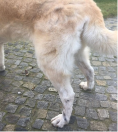
On May 5, 2017, the limbs were measured in a standing position in order to assess the effects of the therapy. The left limb (sick) – thigh circumference = 34 cm, above the knee: 26 cm; the right limb (healthy) – thigh circumference = 39.5 cm, above the knee: 27.5 cm. The described atrophy of the thigh muscles in the left pelvic limb is shown in Figures 1 and 2. Twenty-four days later, another measurement of the left limb was taken – the thigh: 36 cm, above the knee: 27 cm, and this proved the rebuilding of the muscle mass. The measurement of muscle circumference is one of the simplest methods to assess the progress of treatment and recovery.
|
Fig. 1. From the side, left limb |
On May 5, 2017, the last rehabilitation session took place. The dog moved smoothly, it did not limp, the mobility of the joint was not limited, and the muscle mass of the limb was fully rebuilt. Two months later, during the follow-up visit, no features of lameness were reported, the mobility of the operated joint was normal, and there was no visible muscular atrophy.
The rehabilitation sessions were held 2–3 times a week. There were in total 20 therapeutic sessions. Moreover, the owners received a set of home exercises which included: passive exercises of all joints of the operated limb – 20 repetitions 2–3 times a day; Cavaletti hurdles 2 times a day (5 repetitions), and sit-up exercises twice a day (5 repetitions). In addition to the exercises mentioned above, the owners were informed about the positive influence of walking on variable ground and various elevations. As appears from the description, this time the treatment was uneventful, and this is the most common therapy pattern we use in the clinic.
|
Fig. 2. From the back – difference in the thigh circumference is still visible |
On August 14, 2017, the bitch Hovaward, aged 7 years, weighing 35 kg, after an operation on a ruptured cranial cruciate ligament in the left stifle performed on August 11, 2017, was admitted to the clinic. The medical history revealed that the TTA Porus surgical treatment was applied in the university hospital. In this method, a more porous titanium cage is used. Thanks to this solution it is better filled with the new bone. The clinical examination showed that the range of motion in the knee was preserved, there were small muscular atrophies in the thigh, and slight congestive swelling within the ankle. The dog mildly burdened the limb. As physiotherapy was immediately introduced after the surgical procedure the convalescence period was short.
Physiotherapeutic treatments included a constant frequency magnetic field of 15 mT · 10–1, a duration of 20 minutes, and laser therapy with the parameters of 5 J · cm–2 and 5 Hz.
Home rehabilitation covered limb massage (stroking and rubbing). The duration of the massage was up to 15 minutes. Passive exercises – 20 movements per joint and placing the limb on an elevation (e.g. a pillow) in order to reduce congestive swelling. In addition, slow leash walks were recommended to motivate the dog to burden the affected limb. Owners were advised to carry the dog on variable ground. During the day they also had two exercises to choose from Cavaletti hurdles and walking in the figure of eight patterns, 1–2 times a day (20 repetitions).
A total of 15 treatments were performed. The sessions were held 1–2 times a week on average, depending on the time restrictions of the owner.
Three months after the procedure the bitch completely burdened the operated limb, and the muscle mass had fully rebuilt.
The time of convalescence and full recovery after surgery due to RCCL is very diverse, and in the absence of complications, it ranges from 4 to 12 weeks on average. Our observations show that modern tibial osteotomy methods bring better results, and the time to return to full mobility is shorter than in the case of joint stabilization techniques. The rehabilitation plan implemented in the dogs included treatments such as manual therapy, kinesitherapy, and physical therapy. The combination of these rehabilitation methods gives optimal therapeutic effects. In the first dog, the treatment was the longest, i.e. about 6 months, and included about 40 rehabilitation sessions in total. Such a long duration of the therapy resulted from numerous postoperative complications and the need to constantly modify the physiotherapeutic plan and support the patient with pharmacological treatment. It seems that the complications were mostly due to an allergic reaction to the synthetic implant used for extra-capsular stabilization, as well as to the caregivers' failure to observe the physical activity regime after the first treatment, which resulted in implant damage and the need for reoperation. Finally, after long treatment, the dog returned to full mobility. Unfortunately, a year after the end of treatment, walking problems appeared as a result of progressive degenerative knee disease. As in many other complicated cases, this time physiotherapy was also supported by pharmacological treatment ordered by the attending physician. Good cooperation between physiotherapists and physicians is a very important element of therapy, especially when complications occur.
The convalescence of the second patient was faster, and lasted about 3 months; it included 30 therapeutic sessions. This time, a prolongation of the convalescence period may have been due to an initial misdiagnosis and, consequently, a delayed decision to operate. The treatment resulted in the restoration of the full range of motion in the knee joint and regeneration of the muscular system.
The last, third dog recovered the fastest. The medical diagnosis was quick, and 3 days later the dog was operated on. A total of 15 physiotherapy sessions were performed within 4 weeks, followed by a full recovery.
As we can observe, based on the example of the described cases of the three dogs, rehabilitation may have various courses, but it most often leads to regaining the normal motor function of the joint. Therefore, it can be unequivocally concluded that physiotherapy is as equally important as surgery. As in humans, rehabilitation in animals should be a routine procedure. Rupture of the cranial cruciate ligament (CCL) is the most common damage to the dog's knee joint, characterized by muscular atrophy, lameness, and persistent abnormal positioning of the limb [Monk et al. 2006Monk, M.L., Preston, C.A., McGowan, C.M. (2006). Effects of early intensive postoperative physiotherapy on limb function after tibial plateau leveling osteotomy in dogs with deficiency of the cranial cruciate ligament. Am. J. Vet. Res., 67(3), 529–536. https://doi.org/10.2460/ajvr.67.3.529]. Rehabilitation of patients after the operation of the ruptured cranial cruciate ligament is based on the use of anti-inflammatory and analgesic agents, elimination of oedema, maintaining or increasing the range of motion, restoring the muscle mass, and improving proprioception, posture, and gait. Before we start treatment it is very important to know what method was used to stabilize the knee. In the case of TPLO or TTA procedures, in the early stage of rehabilitation, we should take into account proper bone healing [Zink and Van Dyke 2013Zink, M.C., Van Dyke, J.B. (2013). Canine Sports Medicine and Rehabilitation. Wiley‑Blackwell Publishing, Chichester, 1–465. https://doi.org/10.1002/9781118783443], which lasts 8 weeks [Jerram and Walker 2003Jerram, R.M., Walker, A.M. (2003). Cranial cruciate ligament injury in the dog: pathophysiology, diagnosis and treatment. N. Z. Vet. J., 51(4), 149–158. https://doi.org/10.1080/00480169.2003.36357]. In our opinion, it is extremely important to begin postoperative rehabilitation as soon as possible for example by using cold compresses and passive exercises. In each case, it is necessary to consider all indications and contraindications for physiotherapy, resulting from the selected method of surgical treatment and the patient’s condition. The number and duration of the procedures should be modified depending on the healing process progress and possible complications. Other researches also prove that targeted early rehabilitation supports the recovery process in the best possible way and allows a dog to resume normal activity [Romano and Cook 2015Romano, L.S., Cook, J.L. (2015). Safety and functional outcomes associated with short-term rehabilitation therapy in the post-operative management of tibial plateau levelling osteotomy. Can. Vet. J., 56, 942–946. Google Scholar, Baltzer et al. 2018Baltzer, W.I., Smith-Ostrin, S., Warnock, J.J., Ruaux, C.G. (2018). Evaluation of the clinical effects of diet and physical rehabilitation in dogs following tibial plateau leveling osteotomy. J. Am. Vet. Med. Assoc., 252, 686–700. https://doi.org/10.2460/javma.252.6.686, Kirby-Shaw et al. 2020Kirby-Shaw, K., Alvarez L., Tomlinson J.E., Shaw A.J. (2020). Fundamental principles of rehabilitation and musculoskeletal tissue healing. Vet. Surg., 49, 22–32. https://doi.org/10.1111/vsu.13270].
For many years, there have been numerous scientific reports that emphasize the positive impact of rehabilitation as a factor in shortening the recovery period and increasing the chances of regaining full mobility in the operated limb [Marsolais et al. 2002Marsolais, G.S., Dvorak, G., Conzemius, M.G. (2002). Effects of postoperative rehabilitation on limb function after cranial cruciate ligament repair in dogs. J. Am. Vet. Med. Assoc., 220(9), 1325–30. https://doi.org/10.2460/javma.2002.220.1325, Monk et al. 2006Monk, M.L., Preston, C.A., McGowan, C.M. (2006). Effects of early intensive postoperative physiotherapy on limb function after tibial plateau leveling osteotomy in dogs with deficiency of the cranial cruciate ligament. Am. J. Vet. Res., 67(3), 529–536. https://doi.org/10.2460/ajvr.67.3.529, Rexing et al. 2010Rexing, J., Dunning, D., Siegel, A.M., Knap, K., Werbe, B. (2010). Effects of cold compression, bandaging, and microcurrent electrical therapy after cranial cruciate ligament repair in dogs. Vet. Surg., 39(1), 54–58. https://doi.org/10.1111/j.1532-950X.2009.00620.x, Heremans et al. 2017Heremans, J., de Bakker, E., Van Ryssen, B., Samoy, Y. (2017). Therapeutic ultrasound as an aid in tibial fracture management in a dog. Vlaams Diergen. Tijds., 86(1), 29–34. https://doi.org/10.21825/vdt.v86i1.16301]. Rexing et al. [2010]Rexing, J., Dunning, D., Siegel, A.M., Knap, K., Werbe, B. (2010). Effects of cold compression, bandaging, and microcurrent electrical therapy after cranial cruciate ligament repair in dogs. Vet. Surg., 39(1), 54–58. https://doi.org/10.1111/j.1532-950X.2009.00620.x investigated the influence of cold compresses, bandaging, and microcurrent electrical therapy on dogs after extra-capsular stabilization of the ruptured CLL. 24 dogs with CLLR (cranial cruciate ligament rupture) were qualified for the research and divided into 4 study groups. The study showed that bandaging with the so-called Robert Jones bandage was the most effective technique in reducing postoperative swelling of the soft tissues after surgery. However, the best therapeutic effects were obtained when bandaging, cold compresses, and electrotherapy were used in combination. Our assessment indicated that the early post-operative use of cold therapy is particularly important and desirable. Ice compresses definitely reduce pain, swelling, and inflammation after surgery. They should be applied for about 3–4 minutes, but not directly on the skin, so as not to cause tissue frostbite. Optimally, the procedure ought to be repeated every 4 hours. Extensive research on the effects of physiotherapy in the treatment of this disease was conducted by Marsolais et al. [2002]Marsolais, G.S., Dvorak, G., Conzemius, M.G. (2002). Effects of postoperative rehabilitation on limb function after cranial cruciate ligament repair in dogs. J. Am. Vet. Med. Assoc., 220(9), 1325–30. https://doi.org/10.2460/javma.2002.220.1325. The study involved 51 dogs after extra-capsular stabilization of the knee, with a body weight of 20 to 40 kilograms. The animals were divided into 2 groups – the first group consisted of 25 rehabilitated dogs; the other dogs had only instructions given to limit physical activity after the procedure. The rehabilitation included massage, swimming, passive and active exercises, as well as special movement recommendations; the treatments were performed twice a day. In total, each dog underwent 30 treatment sessions within 3 weeks, and the rehabilitation began 2 weeks after the operation. The evaluation of the effects of the therapy was carried out on the basis of measurements of muscle strength and the speed of walking on special floor strength platforms. The measurements of peak vertical force (PVF) and vertical impulse (VI) were taken before and 6 months after surgery. In both groups, after 6 months, the values of PVF and VI increased, but in the rehabilitated group this growth was definitely more significant. More importantly, the values of PVF and VI in the operated limb after 6 months were similar to the outcomes obtained in the healthy limb in dogs undergoing rehabilitation, while in the group not subjected to rehabilitation the function of the operated limb was weaker in comparison to the healthy limb. As we can observe, this time rehabilitation brought significant therapeutic effects. As in our group, the patients were treated with various physiotherapy techniques, which brings the best results. However, it is not always possible to obtain as high a frequency of treatments as in the study described above. Our patients underwent rehabilitation sessions 2–3 times a week and additional home exercises, and this management produced equally positive results. One more conclusion is that in order to objectively evaluate the effect of the treatment, we should focus not only on the assessment of the efficiency of the operated limb but also compare its function with the activity of the healthy limb. It is also extremely important to start rehabilitation as soon as possible after surgery, for example by using cold compresses. Then, the number and time of treatments should be modified depending on the progress of the healing process and possible complications. Heremans et al. [2017]Heremans, J., de Bakker, E., Van Ryssen, B., Samoy, Y. (2017). Therapeutic ultrasound as an aid in tibial fracture management in a dog. Vlaams Diergen. Tijds., 86(1), 29–34. https://doi.org/10.21825/vdt.v86i1.16301 described a case of complicated postoperative physiotherapeutic treatment of the ruptured CCL. A 6-year-old male Bernese Mountain dog with a rupture of the left CCL was subjected to the TTA Rapid procedure. The course of the surgery was uneventful, but 2 weeks later the proximal part of the tibia fractured due to the excessive uncontrolled physical activity of the dog. Conservative treatment included limb stiffening, physiotherapy with ultrasound, and strengthening exercises. As in earlier studies [Claes and Willie 2007Claes, L., Willie, B. (2007). The enhancement of bone regeneration by ultrasound. Prog. Biophys. Mol. Biol., 93, 384–398. https://doi.org/10.1016/j.pbiomolbio.2006.07.021, Mosselmans et al. 2013Mosselmans, L., Samoy, Y., Verleyen, P., Herbots, P., Van Ryssen, B. (2013). Applications of ultrasound in veterinary medicine. Vlaams Diergen. Tijds., 82, 103–111. https://doi.org/10.21825/vdt.v82i3.16702], it has been demonstrated that ultrasound accelerates bone regeneration and adhesion, and increases its endurance, mainly by improving tissue perfusion, and stimulation of protein synthesis, fibroplasia, and collagen production. Only one of the physiotherapeutic methods was used in this patient. In our opinion, better results are obtained by using a variety of treatments, as each of them has a different positive effect on the body. We have distinguished four basic aspects which determine the final effect of the treatment. The first one is a quick diagnosis and surgical treatment because such management minimizes further negative effects of an injury, such as stiffness of the joints, contractures, and muscular atrophy as a result of the non-use of the affected limb. The second aspect involves the choice of the operation method and its proper implementation, with a clear preference for biomechanical methods. The third most important factor concerns complications which are quite often, and usually are, a consequence of surgical errors or improper care of owners after the procedure. The last, fourth aspect concerns the actions of the physiotherapist, namely the choice of the optimal rehabilitation plan and its strict implementation.
In conclusion, the cases we have described, as well as the information mentioned above from the available literature, confirm the effectiveness and thus the need for physiotherapeutic treatment of dogs after the break of CCL. We hope that our physiotherapy treatment regimens will be used in practice in physiotherapy and veterinary practices.
This study was financed by the Educational Fund of the Faculty of Animal Breeding and Biology, Bydgoszcz University of Science and Technology.
Received: 15 Dec 2021
Accepted: 4 Jan 2022
Published online: 2 Jul 2022
Accesses: 1175
Słodki, S., Bogucka, J., (2021). Physiotherapy support for postoperative treatment of cranial cruciate ligament rupture in dogs: case report. Acta Sci. Pol. Zootechnica, 20(4), 25–32. DOI: 10.21005/asp.2021.20.4.04.