Research Article
Anna Wysokińska ![]() , Angelika Chłopik
, Angelika Chłopik
Siedlce University of Natural Sciences and Humanities, Faculty of Agrobioengineering and Animal Sciences, Prusa 14, 08-110 Siedlce, Poland
Abstract. The aim of the present study was to analyze the frequency of occurrence of acrosome-reacted spermatozoa in semen stained with two methods, depending on boar semen storage time. The studies were conducted on ejaculates collected from 10 Landrace boars used in artificial insemination. The smears were prepared by means of two staining methods: SpermBlue and eosin-gentian dye. It was concluded that the proportion of acrosome-reacted spermatozoa depends on the staining method and ejaculate storage duration. When the smears were stained with the SpermBlue method, the number of spermatozoa with head defects, including acrosome reaction, was greater compared with the smears stained with the eosin-gentian dye. With the passage of semen storage time, a change in sperm count with an acrosome reaction was observed. In the SpermBlue-stained smears, the proportion of acrosome-reacted spermatozoa was the highest in the 96th hour of storage and was about 2.5%. An increase in the number of acrosome-reacted spermatozoa was observed in the semen stained with the eosin-gentian dye method in the following semen storage hours.
Keywords: acrosom, boar, semen storage period
Fertilization is a complex multistage process comprising several phases during which spermatozoa undergo structural and metabolic changes. Especially the structure of sperm cell membrane and processes progressing within it during capacitation and acrosome reaction are of crucial significance for the sperm union with oocyte. Acrosome reaction is the last stage of spermatozoa activation. Without it, spermatozoa would not be able to penetrate zona pellucida of ovum. This reaction occurs by the action of acrosomal enzymes released during this process, and prepares spermatozoa for fusion with oolemma. It may be induced by vesicular fluid, chemical compounds (e.g. Ca 2+ ionophore) and progesterone [Tesarik 1985Tesarik, J. (1985). Comparison of acrosome reaction inducing activities of human cumulus oophorus, follicular fluid and ionophore A23187 in human sperm populations of propen fertilizing ability in vitro. J. Reprod. Fert., 74, 383–388. Google Scholar]. For the acrosome reaction to occur, it is essential that sperm membrane integrity is intact and metabolic processes within spermatozoa structures run in a normal way [Šerniené et al. 2005Šerniené, L., Riškevičiené, V., Banys, A., Žilinskas, H. (2005). Comparison of in vitro methods for evaluation of boar semen quality. Vet. Med., 61, 278–281. Google Scholar]. Spermatozoa with premature acrosome reaction are incapable of fusion with egg [Li et al. 2017Li, T., Liu, W., Xie, N., Yang, S.J., Zhang, C., Fu, H.L., Gao, X. (2017). Value analysis of sperm spontaneous acrosome reaction in male fertility evaluation. Andrology, 6 (2), 1–5. https://doi.org/10.4172/2167-0250.1000195]. Cell membrane of boar spermatozoa is particularly sensitive to temperature changes due to its specific structure. It contains a lot of polyunsaturated fatty acids which make it especially vulnerable to damage [Cerolini et al. 2000Cerolini, S., Maldjian, A., Surai, P., Noble, R. (2000). Viability, susceptibility to peroxidation and fatty acid composition of boar semen during liquid storage. Anim. Reprod. Sci., 58, 99–111. Google Scholar]. For this reason, spermatozoa are sensitive to oxidative stress caused by an imbalance between reactive oxygen species production and antioxidant capacity [Agarwal et al. 2008Agarwal, A., Makker, K., Sharma, R. (2008). Clinical relevance of oxidative stress in male factor infertility: an update. Am. J. Reprod. Immun., 59, 2–11. https://doi.org/10.1111/j.1600-0897.2007.00559.x]. When semen is cooled, lipid phase is separated which increases permeability of the sperm cell membrane. Waterhouse et al. [2006]Waterhouse, K.E., Hofmo, P.O., Tverdal, A., Miller, J.R. (2006). Within and between breed differences in freezing tolerance and plasma membrane fatty acid composition of boar sperm. Reproduction, 131, 887–894. https://doi.org/10.1530/rep.1.01049 demonstrated variability of cell membrane composition of spermatozoa produced by different boars, especially in the contents of individual unsaturated fatty acids and cholesterol. Due to these differences, spermatozoa of some service boars are more resistant to techniques of semen processing compared with other AI boars. For these reasons, in assessment of spermatozoa morphology, it is worth taking notice of the condition of cell membrane of boar spermatozoa heads because semen with apparently correct morphological structure can contain spermatozoa with premature acrosome reaction. In artificial insemination (AI) practice, the changes in spermatozoa cell membrane that may occur during storage usually are not analyzed. Routine semen analysis also does not include evaluation of spermatozoa propensity for premature acrosome reaction.
The aim of the present study was to analyze the frequency of occurrence of acrosome-reacted spermatozoa in semen stained with two methods, depending on boar semen storage time.
The studies were conducted on 100 ejaculates collected from 10 Landrace boars used in artificial insemination. Boars selected for the study were 1.5–2 years of age, healthy and maintained at identical environmental conditions. According to the farm standards, the boars were fed a complete diet to meet all of their nutritional needs and had access to water ad libitum. 10 ejaculates per boar were collected at weekly intervals. Ejaculates were collected using a manual method during morning hours at the location of boar use. The gel fraction of semen was removed by gauze filtration. Immediately after collection, volume and spermatozoa count and motility were determined for each ejaculate. Ejaculates chosen for the studies contained not less than 70% of spermatozoa with normal motility. Subsequently, the ejaculates under study were diluted with a commercially available solvent Biosolvens plus (Biochefa, Poland) and insemination doses (closed plastic bags, 100 mL) were prepared, which contained ca. 3 billion spermatozoa in each. Artificial insemination doses prepared in this way were stored in thermobox at 17°C.
Table
1. The frequency of
occurrence of spermatozoa with normal and abnormal morphological
structure depending on the staining method (mean ± SEM) |
||
Item |
Staining metod – Metoda barwienia |
|
SpermBlue |
Eosin-gentian dye |
|
Spermatozoa with normal morphology
|
95.98 ±0.47 |
95.95 ±0.59 |
Spermatozoa with head defects, including: |
2.05a ±0.35 |
1.13b ±0.17 |
– spermatozoa with acrosome reaction |
1.58a ±0.35 |
0.67b ±0.14 |
Spermatozoa with cytoplasmic droplets |
0.39 ±0.08 |
0.32 ±0.11 |
Spermatozoa with tail defects |
1.58a ±0.19 |
2.60b ±0.41 |
a, b – Differences between average values represented by
different letters in the same row (P ≤ 0.05) |
||
Table
2. Coefficients of
phenotypic correlation between spermatozoa with morphological
defects and acrosome-reacted spermatozoa (*P
<
0.05) |
|||
Item |
Spermatozoa with head defects |
Spermatozoa with cytoplasmic droplets |
Spermatozoa with tail defects |
Spermatozoa with acrosome reaction (SpermBlue dye staining) |
0.96* |
–0.07 |
0.26 |
Spermatozoa with acrosome reaction (eosin-gentian dye staining ) |
|
|
|
Samples collected from each artificial insemination dose were used for preparation of semen smears for determination of spermatozoa morphology. The smears were prepared at the following intervals: 1 h, 24 h, 96 h and 168 h after collection.
At each of these times, fresh AI doses were opened immediately before smear preparation. From each insemination dose, two separate samples for microscopic characterization were prepared using different staining methods. In total, 1000 semen smears were prepared.
Eosin-gentian dye staining. A thin semen smear was prepared on defatted microscopic slide warmed to a temperature of 36°C. After drying for ca. 5 min the smear was fixed in 96% ethanol solution. The fixed smears were rinsed with distilled water and stained with 10% aqueous solution of eosin blue (Carl Roth Gmbh+Co. KG, Karlsruhe, Germany) for 20–60 s. The stained smears were rinsed again with water and stained with gentian dye (composed of: 2 g methylene blue, 0.75 g gentian violet, 5 ml glycerol, 100 ml distilled water) (Sigma-Aldrich, USA) for 3–5 min. The smears were rinsed with distilled water and air dried. This procedure led to a clean background and thus, a good contrast against the stained spermatozoa. The slides were prepared and assessed at the same time and by the same person using a microscope.
SpermBlue staining. A 10–15 µl drop of semen was transferred onto a microscopic slide, smeared and air dried at a room temperature. After drying, the smear was stained with the SpermBlue dye (Microptic SL, Spain) for ca. 4 min. Next, the smear was rinsed with distilled water and air dried.
Sperm cell morphology was evaluated under immersion at a magnification of 1000 × with the use of a light microscope Nikon E-50i. In each smear, the morphology of 500 spermatozoa was evaluated by counting spermatozoa with normal and abnormal morphology, according to Blom’s classification [Blom 1981Blom, E. (1981). The morphological estimation of the spermatozoa defects of bull II. The proposal of new classification of spermatozoa defects. Vet. Med., 37, 239–242. Google Scholar].
Morphological defects of spermatozoa were classified into three subgroups:
Experimental data were analyzed using a program STATISTICA 13.1 PL (StatSoft, Tulsa, USA). All results are expressed as mean ±standard error of the mean (SEM). The obtained material was statistically analysed according to the following mathematical model:
The significance of the differences between the groups was assessed with the Tukey test at P ≤ 0.05. Correlations between the acrosome-reacted spermatozoa analyzed with the use of the SpermBlue and the eosin-gentian dye staining methods were calculated by means of the Spearman's rank correlation coefficient with P ≤ 0.05 as the significance level.
Table 1 presents data characterizing the frequency of occurrence of spermatozoa with normal structure and with morphological defects, obtained with the use of two smear staining methods. The data shown in Table 1 indicate that there are differences in the numbers of spermatozoa with morphological defects between semen smears stained with different methods (Fig. 1). When the smears were stained with the SpermBlue method, the number of spermatozoa with head defects, including acrosome reaction, was by 0.92% greater compared with the smears stained with the eosin-gentian dye (P ≤ 0.05). The smears stained with the eosin-gentian dye showed more spermatozoa with tail defects by 1.10% than the SpermBlue-stained smears (P ≤ 0.05).
Table 2 presents correlations between the proportion of morphologically defective spermatozoa and acrosome-reacted spermatozoa stained by the SpermBlue method and the eosin-gentian dye. It was evidenced that there were significant relations between proportion of acrosome-reacted spermatozoa and head defects in smears stained with both methods. Positive significant relations were observed between the number of acrosome-reacted spermatozoa and spermatozoa with cytoplasmic droplets when the smears were stained with the eosin-gentian dye (P ≤ 0.05). The remaining relations proved to be slight and non-significant aspects.
Fig. 1. Eosin–gentian (A–D) and SpermBlue dye staining (E–H): A, E – spermatozoa with normal morphology, B, F – spermatozoa with acrosome reaction, C, G – spermatozoa with a distal droplet, D, H – spermatozoa with a single tail loop
Rys. 1. Metody barwienia eozyna – barwnik gencjanowy (A–D) i SpermBlue (E–H): A, E – plemniki o prawidłowej budowie morfologicznej, B, F – plemniki z reakcją akrosomalną, C, G – plemniki z kroplami distalnymi, D, H – plemniki z pojedynczą pętlą witki
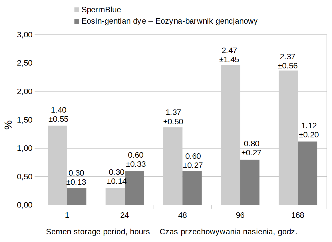
The data presented in Fig. 2 indicate that the proportion of acrosome-reacted spermatozoa increased with semen storage time independently of the smear staining method. The increase in the number of acrosome-reacted spermatozoa with time was more intense in smears stained with the SpermBlue method than with the eosin-gentian dye. In the SpermBlue-stained smears, the proportion of acrosome-reacted spermatozoa rose till the 96th hour of storage of the diluted ejaculate. The percentage of such spermatozoa in the SpermBlue- and eosin-gentian dye-stained smears was 2.37% and 1.12%, respectively. The eosin-gentian dye-stained smears contained a lesser proportion of acrosome-reacted spermatozoa at every hour of semen storage compared with the SpermBlue-stained smears. In the SpermBlue-stained smears, the proportion of acrosome-reacted spermatozoa after 1-hour storage was greater vs. semen stained with the eosin-gentian dye. In SpermBlue-stained smears, the proportion of acrosome-reacted spermatozoa after 1 h and 48 h of storage amounted to 1.40% while after 96 h the proportion rose to almost 2.50%. On the other hand, in the eosin-gentian dye-stained smears, the proportion of acrosome-reacted spermatozoa was at a similar level (0.30–0.60%) over the first two days of storage and gradually rose from 96 h.
Table 3 shows coefficients of phenotypic correlation between acrosome-reacted spermatozoa stained with the SpermBlue method and the eosin-gentian dye. Highly significant positive relations were found between the acrosome-reacted spermatozoa stained with the SpermBlue method after 24, 48, 96 and 168 h of storage and spermatozoa stained with the eosin-gentian dye after 1 h , and after 48 and 168 h of storage.
Table
3. Coefficients of
phenotypic correlation between acrosome-reacted spermatozoa
analyzed with the use of SpermBlue and eosin-gentian dye
staining methods (*P <
0.05) |
||||||
|
SpermBlue |
|||||
1 h |
0.52 |
0.81* |
0.77* |
0.76* |
||
Eosin-gentian dye |
1 h |
0.52 |
0.81* |
0.77* |
0.76* |
0.92* |
24 h |
–0.31 |
–0.20 |
–0.33 |
–0.20 |
–0.37 |
|
48 h |
0.49 |
0.87* |
0.78* |
0.65 |
0.84* |
|
96 h |
–0.01 |
0.44 |
0.24 |
0.19 |
0.55 |
|
168 h |
0.29 |
0.76* |
0.51 |
0.47 |
0.87* |
|
Evaluation of sperm cell head membrane and its sensitivity to premature acrosome reaction can be helpful in assessment of AI boar semen quality. This assessment can be influenced by several factors, including staining solution, smear staining procedure, conditions and duration of semen storage. The data presented in this paper indicate that the smear staining method can affect the result of sperm morphology evaluation. Specifically, semen smears stained with the SpermBlue method were observed to contain significantly less spermatozoa with tail defects but more spermatozoa with head defects, including acrosome reaction, than when the eosin-gentian dye was used for smear staining. Therefore, smear processing procedure can influence the results of sperm morphology evaluation. Staining with the SpermBlue and the eosin-gentian dye produces different results of sperm morphology evaluation in domestic pig males. The SpermBlue method is a relatively new procedure introduced and described in detail by Van der Horst and Maree [2009]Van der Horst, G., Maree, L. (2009). SpermBlue®: A new universal stain for human and animal sperm which is also amenable to automated sperm morphology analysis. Biot. Histoch., 84, 299–308. https://doi.org/10.3109/10520290902984274. Investigations carried out by these authors demonstrated that the SpermBlue staining did not impair sperm head structure of different animal species and humans. The present studies showed that the smears stained with this method contained a higher percentage of spermatozoa with head defects, including acrosome reaction, than after the eosin-gentian dye staining. Considering the present results and data obtained by Van der Horst and Maree [2009]Van der Horst, G., Maree, L. (2009). SpermBlue®: A new universal stain for human and animal sperm which is also amenable to automated sperm morphology analysis. Biot. Histoch., 84, 299–308. https://doi.org/10.3109/10520290902984274, the SpermBlue staining method can be efficient in diagnosing spermatozoa with acrosome reaction. The eosin-gentian dye staining has been commonly used since many years for sperm morphology evaluation of different animal species [Banaszewska et al. 2007Banaszewska, D., Kondracki, S., Wysokińska, A. (2007). The effect of season on the conversion of the morphological structure of sperm insemination boars selected races [Wpływ sezonu na zmiany w budowie morfologicznej plemników wybranych ras knurów inseminacyjnych]. Acta Sci. Pol. Zootech., 6(2), 3–14 [in Polish]. Google Scholar, Kondracki et al. 2012Kondracki, S., Iwanina, M., Wysokińska, A., Huszno, M. (2012). Comparative analysis of Duroc and Pietrain boar sperm morphology. Acta Vet. Brno, 81, 195–199. Google Scholar, Łącka et al. 2016Łącka, K., Kondracki, S., Iwanina, M., Wysokińska, A. (2016). Assessment of stallion semen morphology using two different staining methods, microscopic techniques, and sample sizes. J. Vet. Res., 60 (1), 99–104. https://doi.org/10.1515/jvetres-2016-0014]. Analysis of the smears stained with this method in our studies indicates that spermatozoa are better distinguishable, more intensely stained and there is a greater contrast between spermatozoa and background than in the SpermBlue method. A good contrast in semen smears makes their evaluation easier [Daub et al. 2016Daub, L., Geyer, A., Reese, S., Braun, J., Otzdorff, C. (2016). Sperm membrane integrity in fresh and frozen-thawed canine semen samples: a comparison of vital stains with the NucleoCounter SP-100. Theriogenology, 86, 651–656. https://doi.org/10.1016/j.theriogenology.2016.02.021 !!!]. Daub et al. [2016]Daub, L., Geyer, A., Reese, S., Braun, J., Otzdorff, C. (2016). Sperm membrane integrity in fresh and frozen-thawed canine semen samples: a comparison of vital stains with the NucleoCounter SP-100. Theriogenology, 86, 651–656. https://doi.org/10.1016/j.theriogenology.2016.02.021 !!! recommend analyzing the stained smears shortly after completion of preparation procedure.
Fig. 2. The frequency of occurrence of acrosome-reacted spermatozoa depending on storage duration and smear staining method (mean ±SEM)
Rys. 2. Częstość występowania plemników z reakcją akrosomalną w zależności od czasu przechowywania nasienia i metody barwienia preparatów (średnia ±SEM)
In practice, staining methods create entirely different opportunities for semen diagnosis in different animal species. It was observed that some of them could have adverse effects on the studied cells [Sancho et al. 1998Sancho, M., Perez-Sanchez, F., Tablado, L., de Monserrat, J.J., Soler, C. (1998). Computer assisted morphometric analysis of ram sperm heads: evaluation of different fixative techniques. Theriogenology, 50, 27–37. https://doi.org/10.1016/s0093-691x(98)00110-1, Banaszewska et al. 2015Banaszewska, D., Andraszek, K., Szostek, M., Danielewicz, A., Wójcik, E., Walczak-Jędrzejewska, R. (2015). Analysis of stallion semiologic semen parametrs. Med. Weter., 71, 563–567. Google Scholar]. Some opinions indicate that differences in intensity and contrast of cells stained with various methods can impact on the final result of sperm morphology evaluation [Coetzee et al. 2001Coetzee, K., Bermes, N., Krause, W., Menkveld, R. (2001). Comparison of normal sperm morphology outcomes from two different computer-assisted semen analysis systems. Andrologia, 33, 159–163. https://doi.org/10.1046/j.1439-0272.2001.00421.x, Czubaszek et al. 2019Czubaszek, M., Andraszek, K., Banaszewska, D., Walczak-Jędrzejewska, R. (2019). The effect of the staining technique on morphological and morphometric parameters of boar sperm. PLoS ONE 14 (3), 1–17. https://doi.org/10.1371/journal.pone.0214243]. It is important to choose an appropriate smear staining method in order to minimize its interference with the stained cells [Maree et al. 2010Maree, L., du Plessis, S.S., Menkveld, R., van der Horst, G. (2010). Morphometric dimensions of the human sperm head depend on the staining method used. Hum. Reprod., 25 (6), 1369–1382. https://doi.org/10.1093/humrep/deq075]. According to our observations, a greater contrast between the stained cells and background was achieved after the eosin-gentian dye staining. The use of different dyes may have consequences in relation to evaluation of different spermatozoa structures [Graves et al. 2005Graves, J.E., Lee, H., Boone, W.R., Blackhurt, D.W. (2005). Developing techniques for determining sperm morphology in today’s andrology laboratory. J. Assist. Reprod. Genet., 22, 219–225. https://doi.org/10.1007/s10815-005-4925-3]. Application of dyes with different pH values, osmolarity and staining duration can affect the result of semen morphology evaluation. Studies on semen of Arabian purebred stallions demonstrated that the choice of either eosin-nigrosin or eosin-gentian dye staining did not significantly affect the outcome of evaluation of frequency of morphological sperm defects and that both these methods could be recommended for assessment of stallion sperm morphology [Łącka et al. 2016Łącka, K., Kondracki, S., Iwanina, M., Wysokińska, A. (2016). Assessment of stallion semen morphology using two different staining methods, microscopic techniques, and sample sizes. J. Vet. Res., 60 (1), 99–104. https://doi.org/10.1515/jvetres-2016-0014].
However, it was evidenced that spermatozoa of different animal species showed a variable sensitivity to the same fixatives and staining reagents [Hidalgo et al. 2006Hidalgo, M., Rodriguez, I., Dorato, J. (2006). Influence of staining and sampling procedures on goat sperm morphometry using the Sperm Class Analyzer. Theriogenology, 66, 996–1003. https://doi.org/10.1016/j.theriogenology.2006.02.039, Łukaszewicz et al. 2008Łukaszewicz, E., Jerysz, A., Partyka, A., Siudzińska, A. (2008). Efficacy of evaluation of rooster sperm morphology using different staining methods. Res. Vet. Sci., 85, 583–588. https://doi.org/10.1016/j.rvsc.2008.03.010]. Boar spermatozoa are particularly sensitive due to specific composition of their cell membrane containing higher amounts of polyunsaturated fatty acids [Cerolini et al. 2000Cerolini, S., Maldjian, A., Surai, P., Noble, R. (2000). Viability, susceptibility to peroxidation and fatty acid composition of boar semen during liquid storage. Anim. Reprod. Sci., 58, 99–111. Google Scholar]. Sperm cell membrane is directly exposed to temperature changes in the so-called cold shock range [Kim et al. 2011Kim, S., Lee, Y.J., Kim, Y.J. (2011). Changes in sperm membrane and ROS following cryopreservation of liquid boar semen stored at 15°C. Anim. Reprod. Sci., 124, 118–124. https://doi.org/10.1016/j.anireprosci.2011.01.014, Gączarzewicz et al. 2015Gączarzewicz, D., Udała, J., Piasecka, M., Błaszczyk, B., Stankiewicz, T. (2015). Storage temperature of boar semen and its relationship to changes in sperm plasma membrane integrity, mitochondrial membrane potential, and oxidoreductive capability. Turk. J. Biol., 39, 582–594. https://doi.org/10.3906/biy-1412-76) and to storage conditions and duration [Waberski et al. 2011Waberski, D., Henning, H., Petrunkina, A.M. (2011). Assessment of storage effects in liquid preserved boar semen. Reprod. Dom. Anim., 46, 45–48. https://doi.org/10.1111/j.1439-0531.2011.01836.x]. For this reason, semen should be stored at a temperature above 12°C but not higher than 20°C [Shimatsu et al. 2002Shimatsu, Y., Uchida, M., Niki, R., Imai, H. (2002). Liquid storage of miniature boar semen. Exp. Anim., 51, 143–147. https://doi.org/10.1538/expanim.51.143, Gadea 2003Gadea, J. (2003). Semen extenders used in the artifical insemination of swine. Span. J. Agric. Res., 1, 17–27. Google Scholar]. Ejaculates examined in this study were stored at a temperature of 17°C, i.e. the temperature recommended by Paulenz et al. [2000]Paulenz, H., Kommisrud, E., Homo, P.O. (2000). Effect of long-term storage at different temperatures on the quality of liquid boar semen. Reprod. Dom. Anim., 35, 83–87. Google Scholar, Fantinati et al. [2009]Fantinati, P., Zannoni, A., Bernardini, C., Forni, M., Tattini, A., Seren, E., Bacci, M.L. (2009). Evaluation of swine fertilisation medium (SFM) efficiency in preserving spermatozoa quality during long-term storage in comparison to four commercial swine extenders. Animal, 3, 269–274. https://doi.org/10.1017/S1751731108003443 as optimal for boar semen which should range from 15–20°C. However, semen quality gradually deteriorates with prolongation of storage time [Dziekońska et al. 2013Dziekońska, A., Fraser, L., Majewska, A., Lecewicz, M., Zasiadczyk, Ł., Kordan, W. (2013). Effect of commercial long-term extenders on metabolic activity and membranę integrity of boar spermatozoa stored at 17oC. Pol. J. Vet. Sci., 16, 517–525. https://doi.org/10.2478/pjvs-2013-0072, Wysokińska and Kondracki 2014Wysokińska, A., Kondracki, S. (2014). Assessment of changes in sperm cell membrane integrity occurring during the storage of semen from genetically different males using two diagnostic methods. Can. J. Anim. Sci., 94, 601–606. https://doi.org/10.4141/cjas2013-095, Wysokińska et al. 2015Wysokińska, A., Kondracki, S., Iwanina, M. (2015). The usefulness of selected physicochemical indices, cell membrane integrity and sperm chromatin structure in assessments of boar semen sensitivity. Asian-Austral. J. Anim. Sci., 28 (12), 1713–1720. https://doi.org/10.5713/ajas.15.0095]. Spermatozoa belong to the most highly differentiated cells of the mammalian body and are characterized by high sensitivity to exposure to external factors. Ejaculate processing after collection can generate changes in sperm cellular structures and thus can influence their survival and fertilization ability. In artificial insemination practice, the changes that might occur in the semen after collection and dilution usually are not analyzed. However, factors operating during dilution, preservation and storage of diluted semen can produce structural changes in spermatozoa [Gączarzewicz et al. 2010Gączarzewicz, D., Piasecka, M., Udała, J., Błaszczyk, B., Stankiewicz, T., Laszczyńska, M. (2010). Plasma membrane changes during the liquid storage of boar spermatozoa: a comparison of methods. Acta Vet. Hung., 58, 105–116. https://doi.org/10.1556/AVet.58.2010.1.11, Henning et al. 2012Henning, H., Petrunkina, A.M., Harrison, R.A.P., Waberski, D. (2012). Bivalent response to long-term storage in liquid-preserved boar semen: A flow cytometric analysis. Cytometry Part A., 81(7), 576–587. https://doi.org/10.1002/cyto.a.22058]. Cooling of semen can significantly alter sperm lipid fraction, increase membrane permeability, reduce enzyme activities and change membrane proteins [De Leeuw et al. 1990De Leeuw, F., Colenbrander, B., Verkleij, A. (1990). The role membrane damage plays in cold shock and freezing injury. Reprod. Dom. Anim., 1, 95–104. Google Scholar]. An important component of sperm membrane lipid fraction, cholesterol, is vital for cell membrane permeability. Semen cooling causes cholesterol loss [Cerolini et al. 2001] with concomitant reduction of the stability of sperm cell membrane [Tulsiani et al. 1997Tulsiani, D.R., Yoshida-Komiya, H., Araki, Y. (1997). Mammalian fertilization: a carbohydrate-mediated event. Biol. Reprod., 57, 487–494. Google Scholar]. The present study demonstrated a progressive increase in the proportion of acrosome-reacted spermatozoa during semen storage. A more intense increase in the number of acrosome-reacted spermatozoa was observed in the SpermBlue-stained semen than after the eosin-gentian dye staining. However, these increasing tendencies were similar for both staining methods. The proportion of acrosome-reacted spermatozoa persisted at an analogous level for 48 h of storage to gradually increase in later hours. Some opinions suggest that artificial insemination after storing the semen for four days has an influence in decrease in the number of piglets in the litter [Johnson et al. 2000Johnson, L.A., Weitze, K.F., Fiser, P., Maxwell, W.M.C. (2000). Storage of boar semen. Anim. Reprod. Sci., 62, 143–172. https://doi.org/10.1016/S0378-4320(00)00157-3, Waterhouse et al. 2004Waterhouse, K.E., De Angelis, P.M., Haugan, T., Paulenz, H., Hofmo, P.O., Farstad, W. (2004). Effects of in vitro storage time and extender on membrane quality of boar sperm assessed by flow cytometry. Theriogenology, 62, 1638–1651. https://doi.org/10.1016/j.theriogenology.2004.03.001].
In summary, it can be concluded that the proportion of acrosome-reacted spermatozoa depends on the staining method and ejaculate storage duration. Smears stained with the SpermBlue method contain more spermatozoa with head defects, including acrosome reaction, than the smears stained with the eosin-gentian dye. Both the SpermBlue and eosin-gentian dye staining methods reveal similar tendencies of storage time-dependent changes in the proportion of acrosome-reacted spermatozoa. The number of acrosome-reacted spermatozoa remains at a similar level for the first two days of storage and gradually increases in the next hours. Therefore, it seems that beginning from the third day of storage it is necessary to control the sperm quality more often as well as pay special attention to spermatozoa with defective acrosome.
The work has been financially supported within the part of research task no. 5/19/B – UPH Siedlce.
Received: 25 Oct 2019
Accepted: 31 Dec 2019
Published online: 21 Jan 2020
Accesses: 2034
Wysokińska, A., Chłopik, A., (2019). The occurrence of spermatozoa with acrosome reaction in semen of boars depending on staining method and storage duration. Acta Sci. Pol. Zootechnica, 18(4), 51–58. DOI: 10.21005/asp.2019.18.4.07.